

Книги по МРТ КТ на английском языке / MRI for Orthopaedic Surgeons Khanna ed 2010
.pdf200 |
III Lower Extremity |
|
|
References |
20. Vande Berg BC, Poilvache P, Duchateau F, et al. Lesions of the menisci |
|
|
of the knee: value of MR imaging criteria for recognition of unstable |
|
1. Hardaker WT Jr, Garrett WE Jr, Bassett FH III. Evaluation of acute |
lesions. AJR Am J Roentgenol 2001;176:771–776 |
|
traumatic hemarthrosis of the knee joint. South Med J 1990;83:640– 21. Gentili A, Seeger LL, Yao L, Do HM. Anterior cruciate ligament tear: |
|
|
644 |
indirect signs at MR imaging. Radiology 1994;193:835–840 |
|
2. Khanna AJ, Cosgarea AJ, Mont MA, et al. Magnetic resonance imaging |
22. Umans H, Wimpfheimer O, Haramati N, Applbaum YH, Adler M, |
|
of the knee: current techniques and spectrum of disease. J Bone Joint |
Bosco J. Diagnosis of partial tears of the anterior cruciate ligament of |
|
Surg Am 2001;83:128–141 |
the knee: value of MR imaging. AJR Am J Roentgenol 1995;165:893– |
|
3. Cheung LP, Li KC, Hollett MD, Bergman AG, Herfkens RJ. Meniscal |
897 |
|
tears of the knee: accuracy of detection with fast spin-echo MR |
23. Cone RO III. Knee. Section B. Imaging sports-related injuries of the |
|
imaging and arthroscopic correlation in 293 patients. Radiology |
knee. In: DeLee JC, Drez D Jr, Miller MD, eds. DeLee and Drez’s Ortho- |
|
1997;203:508–512 |
paedic Sports Medicine: Principles and Practice. Philadelphia: WB |
|
4. Deutsch AL, Mink JH, Fox JM, et al. Peripheral meniscal tears: MR |
Saunders; 2003:1595–1652 |
|
findings after conservative treatment or arthroscopic repair. Radiol24. Graf BK, Cook DA, De Smet AA, Keene JS. “Bone bruises” on magnetic |
|
|
ogy 1990;176:485–488 |
resonance imaging evaluation of anterior cruciate ligament injuries. |
|
5. Justice WW, Quinn SF. Error patterns in the MR imaging evaluation of |
Am J Sports Med 1993;21:220–223 |
|
menisci of the knee. Radiology 1995;196:617–621 |
25. Robertson PL, Schweitzer ME, Bartolozzi AR, Ugoni A. Anterior cruci- |
|
6. Kaplan PA, Nelson NL, Garvin KL, Brown DE. MR of the knee: the sig- |
ate ligament tears: evaluation of multiple signs with MR imaging. |
|
nificance of high signal in the meniscus that does not clearly extend |
Radiology 1994;193:829–834 |
|
to the surface. AJR Am J Roentgenol 1991;156:333–336 |
26. Tung GA, Davis LM, Wiggins ME, Fadale PD. Tears of the anterior cru- |
|
7. Kornick J, Trefelner E, McCarthy S, Lange R, Lynch K, Jokl P. Meniscal |
ciate ligament: primary and secondary signs at MR imaging. Radiol- |
|
abnormalities in the asymptomatic population at MR imaging. Radi- |
ogy 1993;188:661–667 |
|
ology 1990;177:463–465 |
27. McCauley TR, Moses M, Kier R, Lynch JK, Barton JW, Jokl P. MR diag- |
|
8. Rubin DA, Paletta GA Jr. Current concepts and controversies in menis- |
nosis of tears of anterior cruciate ligament of the knee: importance |
|
cal imaging. Magn Reson Imaging Clin N Am 2000;8:243–270 |
of ancillary findings. AJR Am J Roentgenol 1994;162:115–119 |
|
9. Stoller DW, Martin C, Crues JV III, Kaplan L, Mink JH. Meniscal tears: |
28. Goldman AB, Pavlov H, Rubenstein D. The Segond fracture of the |
|
pathologic correlation with MR imaging. Radiology 1987;163:731– |
proximal tibia: a small avulsion that reflects major ligamentous |
|
735 |
damage. AJR Am J Roentgenol 1988;151:1163–1167 |
|
10. Wright DH, De Smet AA, Norris M. Bucket-handle tears of the medial |
29. Gross ML, Grover JS, Bassett LW, Seeger LL, Finerman GA. Magnetic |
|
and lateral menisci of the knee: value of MR imaging in detecting |
resonance imaging of the posterior cruciate ligament. Clinical use |
|
displaced fragments. AJR Am J Roentgenol 1995;165:621–625 |
to improve diagnostic accuracy. Am J Sports Med 1992;20:732– |
|
11. Kelley EA, Berquist TH. Knee. In: Berquist TH, ed. MRI of the Mus- |
737 |
|
culoskeletal System. Philadelphia: Lippincott Williams & Wilkins; |
30. Sonin AH, Fitzgerald SW, Friedman H, Ho FL, Hendrix RW, Rogers LF. |
|
2006:303–429 |
Posterior cruciate ligament injury: MR imaging diagnosis and pat- |
|
12. De Smet AA, Norris MA, Yandow DR, Quintana FA, Graf BK, Keene JS. |
terns of injury. Radiology 1994;190:455–458 |
|
MR diagnosis of meniscal tears of the knee: importance of high sig31. Sonin AH, Fitzgerald SW, Ho FL, Friedman H, Bresler ME. MR imag- |
|
|
nal in the meniscus that extends to the surface. AJR Am J Roentgenol |
ing of the posterior cruciate ligament: normal, abnormal, and associ- |
|
1993;161:101–107 |
ated injury patterns. Radiographics 1995;15:551–561 |
|
13. Rubin DA. MR imaging of the knee menisci. Radiol Clin North Am |
32. Escobedo EM, Mills WJ, Hunter JC. The “reverse Segond” fracture: as- |
|
1997;35:21–44 |
sociation with a tear of the posterior cruciate ligament and medial |
|
14. Peterfy CG, Janzen DL, Tirman PFJ, van Dijke CF, Pollack M, Genant |
meniscus. AJR Am J Roentgenol 2002;178:979–983 |
|
HK. “Magic-angle” phenomenon: a cause of increased signal in the |
33. Shelbourne KD, Rubinstein RA Jr. Methodist Sports Medicine Center’s |
|
normal lateral meniscus on short-TE MR images of the knee. AJR Am |
experience with acute and chronic isolated posterior cruciate liga- |
|
J Roentgenol 1994;163:149–154 |
ment injuries. Clin Sports Med 1994;13:531–543 |
|
15. Turner DA, Rapoport MI, Erwin WD, McGould M, Silvers RI. Trunca- |
34. O’Donoghue DH. Treatment of acute ligamentous injuries of the knee. |
|
tion artifact: a potential pitfall in MR imaging of the menisci of the |
Orthop Clin North Am 1973;4:617–645 |
|
knee. Radiology 1991;179:629–633 |
35. Yao L, Dungan D, Seeger LL. MR imaging of tibial collateral liga- |
|
16. Tuckman GA, Miller WJ, Remo JW, Fritts HM, Rozansky MI. Radial tears |
ment injury: comparison with clinical examination. Skeletal Radiol |
|
of the menisci: MR findings. AJR Am J Roentgenol 1994;163:395–400 |
1994;23:521–524 |
|
17. Costa CR, Morrison WB, Carrino JA. Medial meniscus extrusion on |
36. D’Amato MJ, Bach BR Jr. Knee. Section J: Anterior cruciate ligament |
|
knee MRI: is extent associated with severity of degeneration or type |
injuries. Part 1: Anterior cruciate ligament reconstruction in the |
|
of tear? AJR Am J Roentgenol 2004;183:17–23 |
adult. In: DeLee JC, Drez D Jr, Miller MD, eds. DeLee and Drez’s Or- |
|
18. Vande Berg BC, Malghem J, Poilvache P, Maldague B, Lecouvet FE. |
thopaedic Sports Medicine: Principles and Practice. Philadelphia: |
|
Meniscal tears with fragments displaced in notch and recesses |
WB Saunders; 2003:2012–2067 |
|
of knee: MR imaging with arthroscopic comparison. Radiology |
37. LaPrade RF, Gilbert TJ, Bollom TS, Wentorf F, Chaljub G. The mag- |
|
2005;234:842–850 |
netic resonance imaging appearance of individual structures of |
|
19. Lecas LK, Helms CA, Kosarek FJ, Garret WE. Inferiorly displaced flap |
the posterolateral knee. A prospective study of normal knees and |
|
tears of the medial meniscus: MR appearance and clinical signifi- |
knees with surgically verified grade III injuries. Am J Sports Med |
|
cance. AJR Am J Roentgenol 2000;174:161–164 |
2000;28:191–199 |

8 The Knee 201
38.Hughston JC, Eilers AF. The role of the posterior oblique ligament in repairs of acute medial (collateral) ligament tears of the knee. J Bone Joint Surg Am 1973;55:923–940
39.Green NE, Allen BL. Vascular injuries associated with dislocation of the knee. J Bone Joint Surg Am 1977;59:236–239
40.Good L, Johnson RJ. The dislocated knee. J Am Acad Orthop Surg 1995;3:284–292
41.Sonin AH, Fitzgerald SW, Bresler ME, Kirsch MD, Ho FL, Friedman H. MR imaging appearance of the extensor mechanism of the knee: functional anatomy and injury patterns. Radiographics 1995;15:367–382
42.Zeiss J, Saddemi SR, Ebraheim NA. MR imaging of the quadriceps tendon: normal layered configuration and its importance in cases of tendon rupture. AJR Am J Roentgenol 1992;159:1031–1034
43.De Smet AA, Best TM. MR imaging of the distribution and location of acute hamstring injuries in athletes. AJR Am J Roentgenol 2000;174:393–399
44.Koulouris G, Connell D. Evaluation of the hamstring muscle complex following acute injury. Skeletal Radiol 2003;32:582–589
45.Connell DA, Schneider-Kolsky ME, Hoving JL, et al. Longitudinal study comparing sonographic and MRI assessments of acute and healing hamstring injuries. AJR Am J Roentgenol 2004;183: 975–984
46.Brossman J, Preidler KW, Daenen B, et al. Imaging of osseous and cartilaginous intraarticular bodies in the knee: comparison of MR imaging and MR arthrography with CT and CT arthrography in cadavers. Radiology 1996;200:509–517
47.Capps GW, Hayes CW. Easily missed injuries around the knee. Radiographics 1994;14:1191–1210
48.Yu JS, Petersilge C, Sartoris DJ, Pathria MN, Resnick D. MR imaging of injuries of the extensor mechanism of the knee. Radiographics 1994;14:541–551
49.Yu JS, Popp JE, Kaeding CC, Lucas J. Correlation of MR imaging and pathologic findings in athletes undergoing surgery for chronic patellar tendinitis. AJR Am J Roentgenol 1995;165:115–118
50.Kato Y, Oshida M, Aizawa S, Saito A, Ryu J. Discoid lateral menisci in Japanese cadaver knees. Mod Rheumatol 2004;14:154–159
51.Rao PS, Rao SK, Paul R. Clinical, radiologic, and arthroscopic assessment of discoid lateral meniscus. Arthroscopy 2001;17:275–277
52.Rohren EM, Kosarek FJ, Helms CA. Discoid lateral meniscus and the frequency of meniscal tears. Skeletal Radiol 2001;30:316– 320
53.Berquist TH. Musculoskeletal Imaging Companion. Philadelphia: Lippincott Williams & Wilkins; 2002
54.Wantanabe M, Takeda S, Ikeuchi H. Atlas of Arthroscopy. Berlin: Springer-Verlag; 1979
55.Monllau JC, León A, Cugat R, Ballester J. Ring-shaped lateral meniscus. Arthroscopy 1998;14:502–504
56.Yamamoto T, Bullough PG. Spontaneous osteonecrosis of the knee: the result of subchondral insu ciency fracture. J Bone Joint Surg Am 2000;82:858–866
57.Ecker ML, Lotke PA. Spontaneous osteonecrosis of the knee. J Am Acad Orthop Surg 1994;2:173–178
58.Rozing PM, Insall J, Bohne WH. Spontaneous osteonecrosis of the knee. J Bone Joint Surg Am 1980;62:2–7
59.Ramnath RR, Kattapuram SV. MR appearance of SONK-like subchondral abnormalities in the adult knee: SONK redefined. Skeletal Radiol 2004;33:575–581
60.Yates PJ, Calder JD, Stranks GJ, Conn KS, Peppercorn D, Thomas NP. Early MRI diagnosis and non-surgical management of spontaneous osteonecrosis of the knee. Knee 2007;14:112–116
61.Vande Berg BC, Gilon R, Malghem J, Lecouvet F, Depresseux G, Houssiau FA. Correlation between baseline femoral neck marrow status and the development of femoral head osteonecrosis in corticosteroidtreated patients: a longitudinal study by MR imaging. Eur J Radiol 2006;58:444–449
62.Millett PJ, Steadman JR. The role of capsular distention in the arthroscopic management of arthrofibrosis of the knee: A technical consideration. Arthroscopy 2001;17:E31
63.Lindgren PG, Willén R. Gastrocnemio-semimembranosus bursa and its relation to the knee joint. I. Anatomy and histology. Acta Radiol Diagn (Stockh) 1977;18:497–512
64.Bancroft LW. MR imaging of infectious processes of the knee. Magn Reson Imaging Clin N Am 2007;15:1–11
65.Kapoor A, Page S, Lavalley M, Gale DR, Felson DT. Magnetic resonance imaging for diagnosing foot osteomyelitis: a meta-analysis. Arch Intern Med 2007;167:125–132
66.Pineda C, Vargas A, Rodríguez AV. Imaging of osteomyelitis: current concepts. Infect Dis Clin North Am 2006;20:789–825
67.Farley TE, Howell SM, Love KF, Wolfe RD, Neumann CH. Meniscal tears: MR and arthrographic findings after arthroscopic repair. Radiology 1991;180:517–522
68.Davis KW, Tuite MJ. MR imaging of the postoperative meniscus of the knee. Semin Musculoskelet Radiol 2002;6:35–45
69.Lim PS, Schweitzer ME, Bhatia M, et al. Repeat tear of postoperative meniscus: potential MR imaging signs. Radiology 1999;210: 183–188
70.White LM, Schweitzer ME, Weishaupt D, Kramer J, Davis A, Marks PH. Diagnosis of recurrent meniscal tears: prospective evaluation of conventional MR imaging, indirect MR arthrography, and direct MR arthrography. Radiology 2002;222:421–429
71.Potter HG, Rodeo SA, Wickiewicz TL, Warren RF. MR imaging of meniscal allografts: correlation with clinical and arthroscopic outcomes. Radiology 1996;198:509–514
72.Brahme SK, Fox JM, Ferkel RD, Friedman MJ, Flannigan BD, Resnick DL. Osteonecrosis of the knee after arthroscopic surgery: diagnosis with MR imaging. Radiology 1991;178:851–853
73.Dandy DJ, Jackson RW. Meniscectomy and chondromalacia of the femoral condyle. J Bone Joint Surg Am 1975;57:1116–1119
74.Cossey AJ, Kalairajah Y, Morcom R, Spriggins AJ. Magnetic resonance imaging evaluation of biodegradable transfemoral fixation used in anterior cruciate ligament reconstruction. Arthroscopy 2006;22:199–204
75.Macarini L, Murrone M, Marini S, Mocci A, Ettorre GC. [MRI in ACL reconstructive surgery with PDLLA bioabsorbable interference screws: evaluation of degradation and osteointegration processes of bioabsorbable screws]. Radiol Med (Torino) 2004;107:47–57
76.Watanabe BM, Howell SM. Arthroscopic findings associated with roof impingement of an anterior cruciate ligament graft. Am J Sports Med 1995;23:616–625

9 The Foot and Ankle
Daniel J. Durand, John A. Carrino, Meena W. Shatby, Ali Moshirfar, and John T. Campbell
■Specialized Pulse Sequences and Imaging Protocols
Indiscriminate use of MRI can confuse the clinical situation by identifying findings that are not necessarily pathologic or that are poorly correlated with clinical symptoms; these findings in turn can lead to unnecessary and costly interventions and can delay proper diagnosis and treatment.1 MRI of the foot and ankle has improved with the development of high-field systems and enhanced surface coil technology. Most centers use a standard screening examination, consisting of an array of specified imaging planes and pulse sequences. For the ankle, the patient is imaged in a supine, feet-first position with the malleoli several centimeters inferior to the center of the coil; images are obtained in the axial, coronal, and sagittal planes relative to the tabletop. Imaging of the foot is performed in so-called oblique planes, which are parallel and perpendicular to the long axis of the metatarsals, and with minimal plantarflexion to accentuate the fat plane between the peroneal tendons, improve visualization of the calcaneofibular ligament, and decrease the magic angle e ect.2
In addition to standard T1-weighted and T2-weighted sequences, fat-suppressed T2-weighted and STIR sequences are extremely sensitive for detecting marrow and soft-tissue pathology by accentuating the increased signal of fluid and edema relative to the surrounding structures. A protondensity pulse sequence is occasionally used, often with fat suppression; it combines characteristics of T1-weighted and T2-weighted images. These sequences are acquired with an FSE technique that produces the desired contrast in a fraction of the time needed for conventional T2-weighted sequences, minimizing patient motion, improving throughput, and providing great benefit for claustrophobic patients. Novel developments such as parallel imaging techniques,3 combined with increased signal-to-noise ratio at higher field strengths, may continue to reduce scan times.
Imaging parameters can also be refined for optimal cartilage visualization (see Chapter 14). Gradient-echo sequences are often used to evaluate ligaments and articular cartilage (see Chapter 14), particularly in the coronal plane, allowing for optimal visualization of the articular cartilage within the mortise joint. However, early reports comparing cartilage
imaging within the ankle at 3.0 T and 1.5 T have suggested that coronal intermediate-weighted FSE sequences result in better images than do fat-suppressed gradient-echo–based sequences at 3.0 T, primarily because of increased artifact at higher field strength on gradient-echo sequences.4 In addition, gradient-echo sequences should be avoided in the presence of indwelling metal hardware because of the technique’s inherent increased artifact (see Chapter 1).
MR arthrography can be performed as a direct or indirect method. Direct MR arthrography is performed by injecting approximately 10 mL of diluted gadolinium contrast agent into the ankle joint from an anterior approach, entering just medial to the tibialis anterior tendon with slight cranial angulation; care should be taken to avoid the course of the dorsalis pedis artery.5 Postinjection imaging sequences typically include fat-suppressed T1-weighted and T2-weighted images. Indirect MR arthrography is performed by injecting gadolinium intravenously and imaging the joint after contrast has been taken up by the synovium and then di used into the joint.
Direct MR arthrography of the ankle can improve visualization of acute and chronic ligamentous injuries, particularly those involving the LCL complex, although arthrography can also be useful for visualizing syndesmotic and deltoid ligament tears. MR arthrography has also been shown to be an accurate technique for visualizing intraarticular pathology in the various ankle impingement syndromes, in di erentiating stage II from stage III osteochondral talar lesions, and in identifying loose bodies and synovial disorders.5 Indirect MR arthrography is limited by the lack of joint distention and by inferior contrast relative to direct arthrography, although it can be a useful adjunct to MRI for the assessment of the following5:
•Subtle cartilage defects
•Osteochondral talar lesions
•Ligamentous injuries
•Synovitis
•Sinus tarsi syndrome
However, with the advent of high-field, high-resolution imaging and novel noncontrast MR techniques, direct and indirect MR arthrography techniques may be rendered obsolete in the future.
202
|
|
9 The Foot and Ankle 203 |
||
|
|
is not necessary for most acute ankle sprains, but it can be |
|
|
■ Traumatic Conditions |
||||
of use in certain cases of atypical presentation or to rule out |
||||
Bone Contusion |
other injuries such as an OCD or tendon injury. Within the |
|||
Bone contusion represents an injury to the internal bone |
first week after an ankle sprain, MRI usually reveals promi- |
|||
nent subcutaneous edema and juxtaarticular hematoma; |
||||
structure with microtrabecular disruption, but no gross corti- |
these findings present as high signal fluid in the ankle joint |
|||
cal fracture. On MRI, bone contusion appears as a focal area of |
and adjacent tendon sheaths on T2-weighted images, indi- |
|||
low signal on T1-weighted images with ill-defined margins; |
cating the presence of blood within these compartments.6,8 |
|||
high signal on T2-weighted images signifies edema within the |
The edema typically diminishes rapidly thereafter, although |
|||
bone marrow but an intact cortical structure6 (Fig. 9.1). Bone |
fascial edema around the injured ligament may persist.6,8 |
|||
bruises may be seen after direct trauma or after acute ankle |
After the first several weeks, the ligament becomes thick- |
|||
sprains.6,7 Certain bone-bruise patterns are characteristic for |
ened and may appear in continuity despite a tear, which may |
|||
specific injury mechanisms. For example, inversion injuries |
confound accurate interpretation. Two months after injury, |
|||
are associated with contusion of the medial malleolus and |
fascial edema is resolved and the torn ligaments may appear |
|||
medial talus (with signal changes seen in those structures), |
intact, thinned, attenuated, or even completely absent, de- |
|||
whereas a plantarflexion mechanism can result in a posterior |
pending on the degree of initial injury.6,8 |
|||
tibial bone bruise. Jamming injuries of the hallux may result |
|
|
||
in a bone contusion of the first metatarsal head dorsally, but |
Lateral Ankle Sprains |
|||
direct impact on the ball of the foot may result in contusion of |
||||
|
|
|||
a sesamoid. Acute bone bruises typically resolve within 2 to 3 |
Ankle sprains are the most common athletic injury, and spe- |
|||
months unless there is ongoing repetitive injury. |
cifically, lateral ankle ligament sprains are the most common |
|||
|
|
type.9 Ankle sprains account for 45% of basketball injuries,10 |
||
Ligament Sprains |
and are commonly seen in football, soccer, rugby, and vol- |
|||
Ankle sprains are an extremely common injury, both during |
leyball players. These injuries vary in severity from mild to |
|||
severe6,10; lateral ligament injuries are more common than |
||||
sports and during daily activities. The vast majority can be |
medial ones.9,10 The most commonly injured ligament is the |
|||
diagnosed with a detailed history, careful physical examina- |
anterior talofibular ligament, followed by a combination |
|||
tion, and ankle radiographs to exclude acute fracture. MRI |
injury of the calcaneofibular and anterior talofibular liga- |
|||
|
|
ments. An isolated posterior talofibular ligament tear is rare. |
||
|
|
The most common mechanism of injury is inversion and in- |
||
|
|
ternal rotation. It must be noted that this mechanism can |
||
|
|
also produce other common injuries, in isolation or in com- |
||
|
|
bination with ankle sprain, such as the following: |
||
• Fibular or talar fractures
• Osteochondral lesions
• Peroneal tendon tears
• Fifth metatarsal fractures
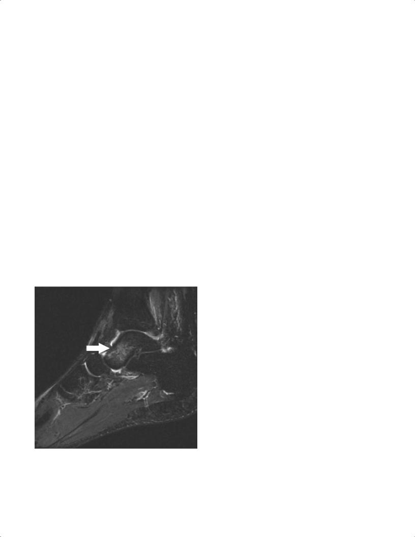
Fig. 9.1 A sagittal T2-weighted image with fat suppression showing a talar contusion. Note the focus of high signal within the talus (arrow), which signifies edema within the bone marrow. The cortex is intact.
The diagnosis of ankle sprain is usually accomplished with a thorough history and physical examination. Conventional radiographs may reveal any associated avulsion fractures or osteochondral injuries, and stress views can be obtained to evaluate structural instability. Occasionally, MRI is used to confirm the diagnosis or for the evaluation of associated injuries. Ligament injuries are best evaluated on T2-weighted and fat-suppressed T2-weighted MR images. These images usually show increased signal intensity that represents the edema and fluid collection secondary to trauma.6,8 Discontinuity of the ligament on T1-weighted and T2-weighted images signifies disruption.6,8 The anterior talofibular ligament is the weakest and the most vulnerable of all ankle ligaments. Tears in this ligament are best diagnosed on axial images (Fig. 9.2).
The second most common pattern of ankle injury is a combined injury to the anterior talofibular ligament and
204 III Lower Extremity
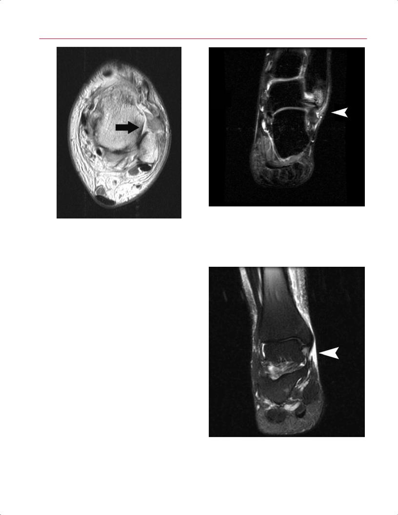
Fig. 9.2 An axial T1-weighted image of the left ankle at the level of the superior talus. No ligament is seen at the expected location of the attachment of the anterior talofibular ligament (arrow). Instead, there are ill-defined fibers extending anteriorly from the fibula, representing a grade III tear.
the calcaneofibular ligament.10 Isolated tears of the calcaneofibular ligament are extremely uncommon.10 Unlike the anterior talofibular ligament, tears of the calcaneofibular ligament are best seen on the coronal T2-weighted, fatsuppressed T2-weighted, and fat-suppressed STIR images (Fig. 9.3). MRI grading of the ligamentous injury can be categorized as follows6:
•Grade I, intact ligament structure but surrounding edema as seen on T2-weighted images
•Grade II, thickening or partially torn fibers of the ligament and surrounding edema
•Grade III, complete disruption of the ligament and loss of its attachment as seen on T1-weighted and T2weighted images
Medial Ankle Sprains
Isolated injuries of the deltoid ligament are uncommon.10 They often occur in conjunction with a fibular fracture or a purely ligamentous syndesmotic injury. Deltoid ligament injuries are best viewed on coronal T2-weighted and fatsuppressed T2-weighted images (Fig. 9.4). Occasionally, medial edema and deltoid thickening are seen on T2-weighted images secondary to an inversion injury.6 This medial
Fig. 9.3 A coronal STIR image of the left ankle showing edema and fluid (arrowhead) tracking longitudinally along the fibers of the calcaneofibular ligament, representing a grade II tear. There is also edema of the distal fibula.
Fig. 9.4 A coronal fat-suppressed T2-weighted image of the right ankle showing a grade II or grade III tear (arrowhead) of the deltoid ligament.
|
9 The Foot and Ankle 205 |
||
|
|
|
|
impaction is usually associated with lateral soft-tissue |
Clinical examination is supplemented with radiographic |
||
edema or evidence of a lateral ligament tear. MR images |
studies to exclude fibular fracture or diastasis of the syn- |
||
must be correlated carefully with clinical examination to |
desmosis. One study has indicated that the measurements |
||
avoid treatment of false-positive findings. |
on static ankle radiographs are poorly predictive of syndes- |
||
|
motic injury.11 Dynamic stress radiographs may identify in- |
||
Syndesmotic Injuries11,12 |
stability of the syndesmosis, but they may be limited in the |
||
acute setting because of patient discomfort. |
|||
|
|||
Also sometimes called high ankle sprains, syndesmotic |
MRI has been found to very useful in identifying these |
||
sprains are reported to occur in as many as 18% of all ankle |
injuries. In one study, MRI had a sensitivity of 100%, a speci- |
||
sprains in athletes.13 Syndesmotic injury usually is associ- |
ficity of 93%, and an accuracy of 97% in diagnosing syndes- |
||
ated with a fibular fracture, but it can occur as a purely liga- |
motic injuries.12 The anteroinferior tibiofibular ligament |
||
mentous diastasis.12,14 The major contributors to the distal |
and posteroinferior tibiofibular ligament are best viewed in |
||
tibiofibular syndesmotic structure are the following: |
axial images at the level of the tibial plafond. When injured, |
||
• Anteroinferior tibiofibular ligament |
the ligaments can be seen as edematous, wavy, absent, or |
||
discontinuous on T1-weighted and T2-weighted sequences |
|||
• Posteroinferior tibiofibular ligament |
|||
(Fig. 9.5A). Extension of fluid into the syndesmosis on axial |
|||
• Interosseous membrane |
|||
T2-weighted images can also assist in the diagnosis of syn- |
|||
|
|||
The anteroinferior tibiofibular ligament is most commonly |
desmotic disruption (Fig. 9.5B). |
||
involved in syndesmotic injuries. External rotation is the |
|
|
|
typical mechanism of injury, and many of these injuries are |
OCD of the Talus |
||
not recognized initially unless the clinician has a high level |
|
|
|
of suspicion for them.13 Patients typically present with se- |
OCD, also known as an osteochondral fragment of the talus, |
||
vere edema, ecchymosis, and tenderness more laterally than |
is a disruption of that structure’s cartilage surface. Analo- |
||
would be expected with a standard lateral ligament sprain. |
gous lesions are common in the knee and elbow. Many occur |
||
A |
B |
||
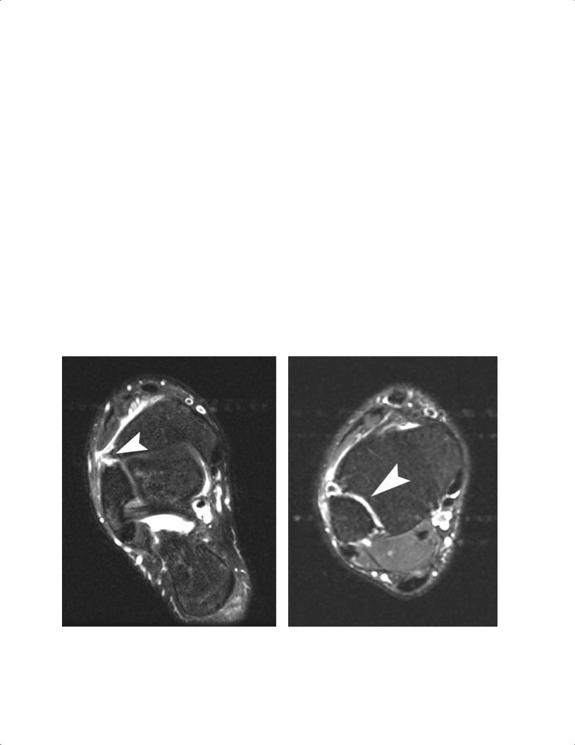
Fig. 9.5 MRI can reveal direct or indirect evidence of syndesmotic injury. (A) An axial fat-suppressed T2-weighted image of the right ankle shows ill-defined, wavy fibers of a torn anteroinferior tibiofibular ligament (arrowhead), direct evidence of a syndesmotic sprain.
(B) More superiorly on the same patient, there is extension of fluid into the syndesmosis (arrowhead), providing indirect evidence of syndesmotic disruption.
206 III Lower Extremity
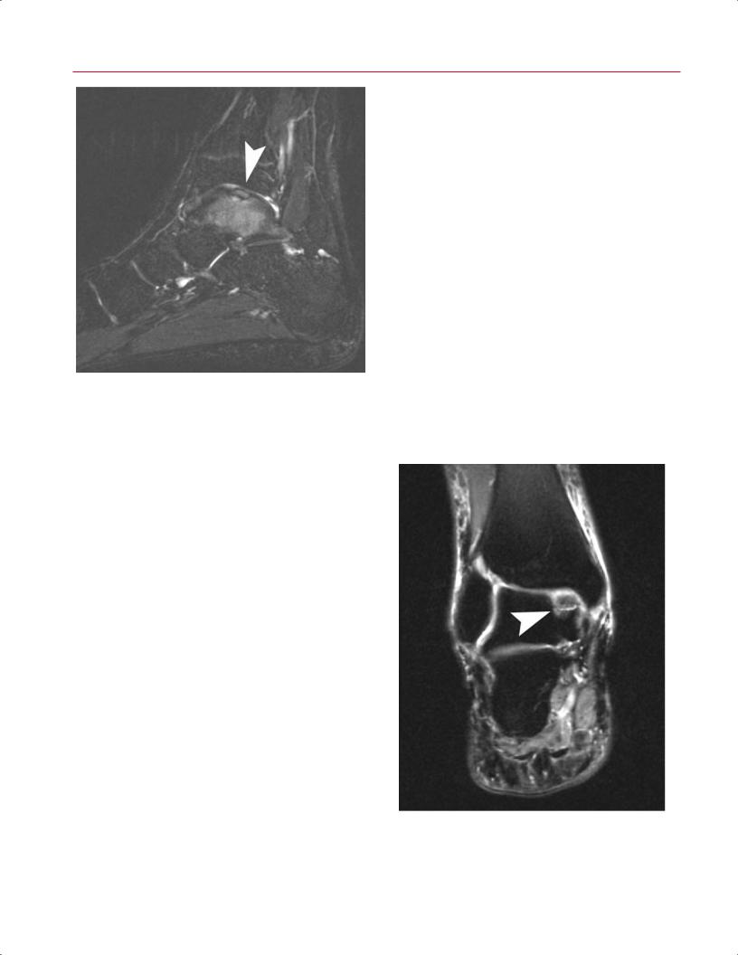
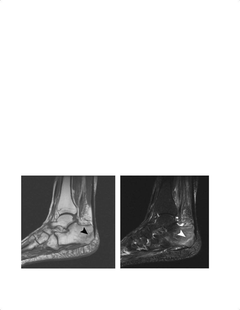
Fig. 9.6 A sagittal fat-suppressed T2-weighted image showing the typical MRI appearance of an OCD, with osseous edema and irregularity of the subchondral bone plate and overlying cartilage (arrowhead).
after a traumatic injury, although some patients do not recall a specific injury. An OCD lesion often is misdiagnosed clinically as an ankle sprain.15,16 Patients typically present with pain located deep within the joint that is worsened with activity or sports; patients may also complain of ankle swelling, giving way, or locking.
OCD lesions can occur anywhere on the surface of the talar dome; the classic literature notes anterolateral and posteromedial lesions as the most common.15,17 A recent MRI study identified the centromedial and centrolateral locations as the most common.18
A spectrum of OCD lesions exists, including the follow-
ing16,19:
•Softening of the cartilage
•Partial detachment of an osteochondral fragment
•Complete detachment of an osteochondral fragment
•Free-floating fragment within the joint
•Subchondral cystic lesion
Stable lesions may respond to nonsurgical treatment, whereas unstable lesions and cystic OCD lesions typically are treated surgically.
MRI is a very useful modality for diagnosing OCD lesions, particularly if the lesion is not visualized on standard radiographs. MRI can also assess other pathologic entities, such as tendon or ligamentous injuries, in the patient with persistent pain after ankle sprain. All three imaging planes
can be useful in assessing OCD lesions. The AP location can be determined on axial and sagittal images, whereas the mediolateral location can be assessed on coronal and axial views. The depth of the lesion can be viewed on sagittal and coronal images. The MRI appearance of an OCD lesion typically shows osseous edema along with irregularity of the subchondral bone plate and overlying cartilage (Fig. 9.6). Specialized cartilage-specific sequences can improve imaging of the chondral surface to identify subtle disruption20 (see the detailed discussion in Chapter 14). In some cases, the surrounding bone edema may cause overestimation of the true size of the lesion. MRI appearance also may assist in predicting the stability of the lesion; fluid or granulation tissue beneath the lesion at the interface with the osseous crater appears bright on T2-weighted imaging and implies instability21–23 (Fig. 9.7). MRI has also been shown to be of use in evaluating healing of the OCD lesion after surgery, with some normalization of signal changes noted.24,25
Stress Fractures
Osseous stress fractures occur from repetitive stress that is below the threshold for acute fracture but substantial enough to cause microtrabecular failure. They often occur
Fig. 9.7 A coronal fat-suppressed T2-weighted image of the right ankle showing an unstable OCD with fluid beneath the lesion at the interface with the osseous crater of the medial talus (arrowhead).
|
9 The Foot and Ankle 207 |
||
|
|
|
|
secondary to overuse situations, such as in an athlete with |
of the heel. Sometimes conventional radiographs can show a |
||
a sudden, dramatic increase in running or other training. |
radiolucent line at the posterior aspect of the calcaneus per- |
||
The bone is unable to heal adequately because of ongo- |
pendicular to the trabecular cancellous lines. MRI is useful if |
||
ing insult, and a stress fracture results. These fractures are |
radiographs are negative, with T1-weighted images showing |
||
typically diagnosed based on a history of repetitive overuse |
a linear fracture line and T2-weighted images revealing as- |
||
and are di erentiated from acute fractures by the lack of |
sociated marrow edema (Fig. 9.8). |
||
an acute traumatic event. The location of lower extremity |
|
|
|
stress fractures in a group of military recruits has been re- |
Navicular Stress Fractures |
||
ported to vary by gender: the most common sites in males |
|||
|
|
||
were the metatarsals (66%), calcaneus (20%), and lower leg |
Navicular stress fractures commonly present in an athlete |
||
(13%), whereas the most common sites in females were the |
who complains of vague arch pain that is worsened with |
||
calcaneus (39%), metatarsals (31%), and lower leg (27%).26 |
running or sports activities. The stress fracture usually oc- |
||
Furthermore, conventional radiographs usually are negative |
curs in the middle third of the navicular secondary to in- |
||
early in the process until callus appears or the stress fracture |
creased stresses in that region along with poor local vascular |
||
progresses to a frank fracture. One study found a rate of posi- |
supply to the bone. MRI typically shows edema within the |
||
tive radiographic findings of only 10% in patients with stress |
navicular on T2-weighted and fat-suppressed T2-weighted |
||
fractures.27 MRI is a very helpful tool in evaluating these in- |
images (Fig. 9.9A). T1-weighted images show a low inten- |
||
juries, with high sensitivity and specificity. Typically, the os- |
sity linear lesion representing a fracture line (Fig. 9.9B). If |
||
seous structures have a linear region of low signal intensity |
MRI suggests a navicular stress fracture, a CT scan may be |
||
on T1-weighted and T2-weighted images; T2-weighted im- |
indicated to define more clearly the osseous structure and |
||
ages also show surrounding high signal intensity consistent |
extent of the fracture. |
||
with bone marrow edema.6 |
|
|
|
Calcaneal Stress Fractures |
Metatarsal Stress Fractures |
||
Metatarsal stress fractures are also called march fractures be- |
|||
|
|||
A patient with a calcaneal stress fracture presents with pos- |
cause they often present in military recruits who participate |
||
terior heel pain and, occasionally, swelling. On physical ex- |
in extended marching drills. Metatarsal stress fractures also |
||
amination, pain is elicited with medial-lateral compression |
commonly present in female dancers who are often in the |
||
A B
Fig. 9.8 Calcaneal stress fracture. (A) A sagittal T1-weighted image showing an oblique fracture line in the posterior calcaneus (arrow-
head). (B) A sagittal fat-suppressed T2-weighted image shows marrow edema and edema tracking along the fracture line (arrowhead).
208 III Lower Extremity
A  B
B
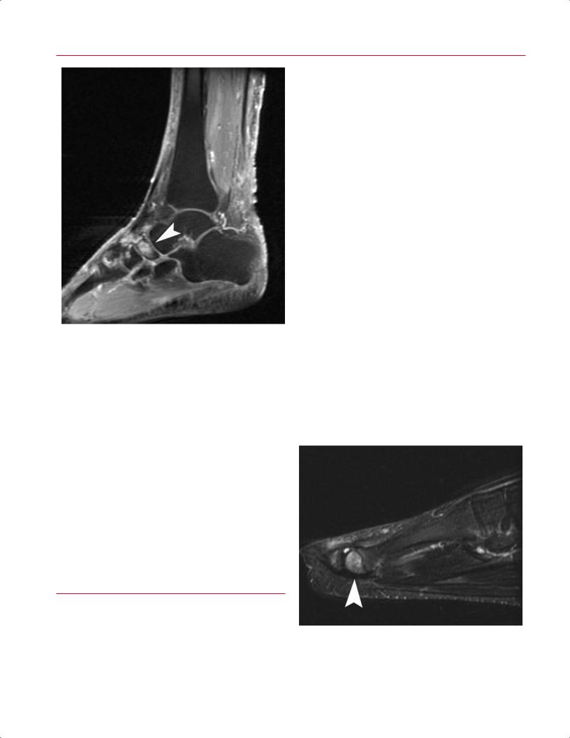
Fig. 9.9 Navicular stress fracture. (A) A sagittal fat-suppressed T2weighted image showing marrow edema (arrowhead) within the navicular. (B) A sagittal T1-weighted image showing a hypointense
en-pointe position and in female athletes, both of whom subject the central metatarsals to increased mechanical load. The second metatarsal is the most frequent location for fracture (more than half of all metatarsal stress fractures), followed by the third and fourth metatarsals.28 Stress fractures of the central metatarsals typically occur in the diaphysis, whereas a stress fracture of the first metatarsal occurs in the proximal metaphysis and a stress fracture of the fifth metatarsal occurs at the diaphyseal-metaphyseal junction.28 Patients typically complain of aching pain related to weight-bearing activities, running, or sports. Conventional radiographs often are diagnostic, although they may be negative early in the process. MRI can be useful in such cases and to rule out other causes of pain such as synovitis. The metatarsal shows the characteristic linear density on T1-weighted images with high signal on corresponding T2-weighted and fat-suppressed T2-weighted images (Fig. 9.10).
■ Degenerative Conditions
Tendon Disorders
region within the dorsal aspect of the navicular with a faint horizontally oriented fracture line (arrowhead).
disorders and is best achieved by obtaining images perpendicular to the tendon’s course.29–33 At the ankle and hindfoot levels, axial views are ideal for tendon imaging, whereas coronal images are best for visualizing more distal structures in
Tendon disorders can represent traumatic injuries, inflammatory conditions, or degenerative tendinosis. Regardless of the etiology, MRI is the ideal method of evaluating tendon
Fig. 9.10 A sagittal fat-suppressed T2-weighted image showing edema of the head of the second metatarsal (arrowhead) in a patient with a stress fracture.
9 The Foot and Ankle 209
•Insertional tendinosis
•Noninsertional tendinosis
•Rupture
Fig. 9.11 Adventitial bursitis as seen on a sagittal fat-suppressed T2weighted image showing soft-tissue inflammation posterior to the calcaneus (arrowhead).
the midfoot. The magic angle e ect is a phenomenon seen in tissues with well-ordered collagen fibers, such as a tendon.34 This phenomenon occurs when the images are obtained with the magnetic field at an oblique angle (55 degrees) relative to the fibers of the tendon, and it shows abnormal signal intensity on T1-weighted images. T2-weighted images are unaffected by this phenomenon, so corresponding T2-weighted images are scrutinized in the a ected areas to determine if the signal abnormality seen on T1-weighted images represents true pathology or simply artifact. The magic angle e ect is seen most notably in the posterior tibialis and peroneal tendons as they course around the malleoli. Knowledge that this magic angle e ect exists, combined with assessment of the T2-weighted images, assists the radiologist or surgeon in accurately identifying pathology of the tendons about the ankle.
Achilles Tendon Disorders
Achilles tendon disorders represent a spectrum of disease, including the following:
•Retrocalcaneal bursitis
•Paratenonitis
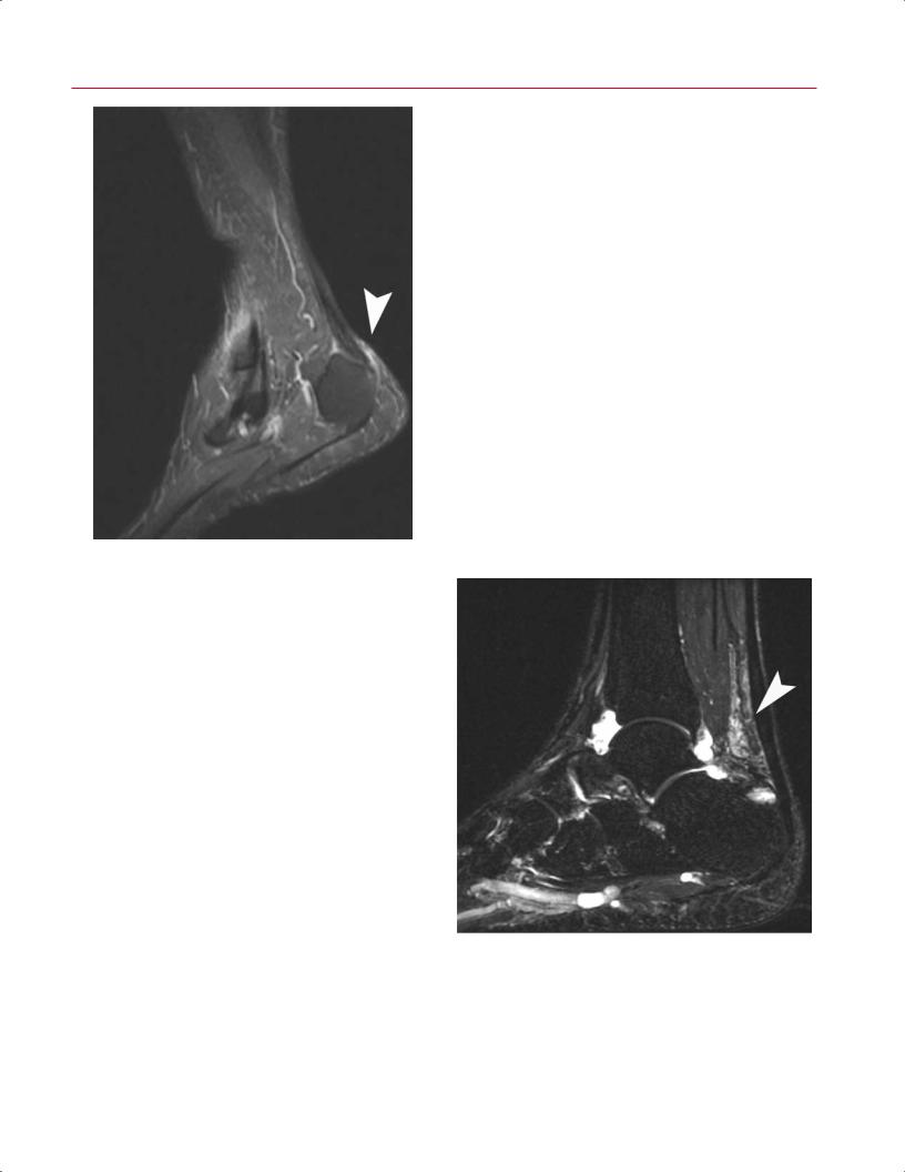
Isolated retrocalcaneal bursitis can be seen secondary to systemic inflammatory disease or localized soft-tissue irritation from impingement by the posterosuperior process of the calcaneus (Haglund process). Sagittal MR images show decreased signal intensity on T1-weighted images and increased intensity on T2-weighted and fat-suppressed T2-weighted images, consistent with soft-tissue inflammation6 (Fig. 9.11). Paratenonitis represents a di use inflammation of the investing paratenon layer that surrounds the Achilles tendon. Sagittal T2-weighted and fat-suppressed T2-weighted images show loss of the normal sharp interface between the Achilles and surrounding soft tissues but normal intrasubstance appearance and morphology of the tendon itself (Fig. 9.12).
The term Achilles tendinitis now is believed to be a misnomer because the tendon typically does not show an inflammatory component. Instead, tendinosis is the preferred term, which more accurately reflects the disorganized collagen fibers, myxoid degeneration, and microscopic intrasubstance tearing seen histologically.35 Achilles tendinosis
Fig. 9.12 The MRI appearance of Achilles paratenonitis is defined by di use inflammation of the investing paratenon layer that surrounds the Achilles tendon. This sagittal fat-suppressed T2-weighted image shows inflammation of the soft tissues surrounding the Achilles (including Kager’s fat pad) (arrowhead) but normal intrasubstance appearance and morphology of the tendon itself. An ankle joint effusion is also seen.