

Книги по МРТ КТ на английском языке / PLUM AND POSNER S DIAGNOSIS OF STUPOR AND COM
.pdf206 Plum and Posner’s Diagnosis of Stupor and Coma
anesthesia, even profound suppression of neural activity resulting from self-administered barbiturates or other sedative drugs. Even when coma is so deep that artificial respiration must be provided for several days and the blood pressure supported by vasopressor agents for a week or more, patients can awaken with no apparent or measurable impairment of brain function. Hence, it is critical to determine the presence of sedative overdose when evaluating the prognosis of a patient in coma, even those with other causes of coma.
The complete reversibility of anesthetic coma, plus the low metabolic rate that accompanies deep anesthesia, has inspired efforts to determine whether barbiturate anesthesia can minimize the expected extent of postanoxic ischemic brain damage. Barbiturates also scavenge free radicals from reoxygenated tissue, but it remains to be proved that this represents an important biologic function in resuscitation. On the other hand, phenobarbital also induces cytochrome P450, which serves as a source of reactive oxygen species. Whether these oppo-
site effects help, hurt, or have no effect on the brain is unclear.104,105 Of some interest, a ran-
domized trial of neonates with hypoxicischemic encephalopathy indicates that phenobarbital in a dose of 20 mg/kg given intravenously within 6 hours of birth in term and near-term neonates was associated with a decrease in lipid peroxides. There was also a decrease in antioxidant enzymes and antioxidant vitamins in the CSF. A trend suggested that lower levels of lipid peroxides in the CSF were associated with a better outcome.106 Barbiturate coma is effective in controlling intractable status epilepticus, but its role in any other brain injury is, at this writing, uncertain.101 Barbiturate anesthesia has been applied to patients in coma from head trauma. It lowers ICP, but it is unclear if it affects outcome.107
MECHANISMS OF IRREVERSIBLE ANOXIC-ISCHEMIC BRAIN DAMAGE
Anoxia, ischemia, and hypoglycemia, although biologically different,108 can combine under several circumstances to damage the brain. Somewhat different but overlapping pathologic changes characterize the irreversible brain injury caused by each of these three
conditions. Systemic and local circulatory differences among them influence the exact geography and type of cellular response. Similar changes in the brain mark the postmortem findings of several conditions, including patients dying in coma after fatal status epilepticus, carbon monoxide poisoning, or several of the systemic metabolic encephalopathies.
Global Ischemia
Complete cerebral ischemia, as in cardiac arrest in man, causes loss of consciousness in less than 20 seconds. Within 5 minutes, glucose and high-energy phosphate stores are depleted. Following that the patient, even if successfully resuscitated, may be left severely brain damaged. This is especially true in elderly patients who most frequently suffer cardiac arrest because their brains are more vulnerable to ischemic damage. By definition, during cardiac arrest the CBF falls to zero. Resuscitation results in transient hyperemia with increased blood flow and oxygen metabolism; subsequently, both decrease in a heterogeneous fashion.109 In most patients, when blood flow is re-established, cerebral autoregulation is either absent or the curve is shifted to the right, such that CBF begins to fall at a higher mean arterial pressure than it did before the cardiac arrest. As a result, it is important to maintain normal and perhaps slightly elevated blood pressure after cardiac arrest.
Both vascular and neuronal factors play a role in the seemingly brief periods of global ischemia that can damage the brain in clinical circumstances. Changes to vascular endothelium during the course of ischemia, as well as additional changes to glial cells (swelling to compress endothelial vessels, viscosity changes in blood), may lead to poor perfusion once cardiac function is restored. This so-called ‘‘no-reflow phenomenon’’110 increases with prolonged duration of ischemia.110–112 Loss of autoregulation can aggravate edema formation, lead to hemorrhage, and cause additional neuronal damage, so-called ‘‘reperfusion injury.’’113 The combination of the ischemia and its aftermath results in neuronal necrosis,114 particularly in the hippocampus, but if the ischemia is prolonged, elsewhere in the hemispheres as well.
Although the exact mechanisms are not understood, it is likely that during the ischemia
Multifocal, Diffuse, and Metabolic Brain Diseases Causing Delirium, Stupor, or Coma |
207 |
the loss of high-energy phosphates causes cellular depolarization that induces the release of glutamate, which in turn causes entry of toxic levels of calcium into neurons. In the reperfusion phase, the restoration of oxidative metabolism probably produces a burst of excess free radicals that are also cytotoxic.113
Cardiac arrest can either cause death of neurons, particularly in vulnerable areas associated with reactive astrocytes, or microinfarcts and areas of pancellular necrosis associated with perivascular diffuse tissue spongiosis. The latter lesions appear in a laminar distribution and are more profound in watershed zones between the major territories of arterial supply. Both types of lesions are more intense and heterogeneous in patients dying after a period of prolonged coma.115
Particularly vulnerable areas include the occipital cortex, the frontoparietal cortex, the hippocampus, the basal ganglia, the thalamic reticular nucleus, Purkinje cells of the cerebellum, and the spinal cord (Figure 5–5). Laminar necrosis of the cortex generally involves layers III and V, which contain the greatest numbers of large pyramidal cells. The most vulnerable area is the CA1 region of the hippocampus. Some patients with lesions restricted to the CA1 region who recover from cardiac arrest can develop a residual severe anterograde amnesia (see Patient 5–6).116
Focal Ischemia
Focal ischemia differs from global ischemia in that it allows for collateral circulation to deliver at least some blood to the areas surrounding the area of no perfusion induced by the vas-
cular occlusion. The surrounding area, called the penumbra,117 suffers low flow but not cel-
lular death. It is the goal of the physician treating the patient to try to preserve that area and return its metabolism to normal. Like global ischemia, damage can occur either during the ischemic period or during reperfusion.118 Schaller and Graf118 have diagramed a threepeaked curve presenting times at which the penumbra is susceptible to tissue damage. The first occurs during ischemia with damage resulting from oxygen depletion, energy failure, depolarization of neurons and synapses, and homeostasis failure. The second occurs after reperfusion with damage caused by excitotox-
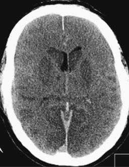
Figure 5–5. Computed tomography scan of a comatose patient after prolonged cardiopulmonary resuscitation. The scan was taken at a time when the patient was deeply comatose but breathing. The brain is swollen, with cortical sulci virtually obliterated. No signal differentiation can be seen between gray and white matter. The lentiform nuclei are hypointense, suggesting basal ganglia infarction. (Scan courtesy Dr. Sasan Karimi.)
icity as well as disturbed homeostasis. The third occurs several weeks later with late damage to neurons and glial cells via both necrosis and apoptosis. As indicated above, interventions that appear to ameliorate the first two peaks, such as the use of anesthetic agents at the time of ischemia, do not appear to have any effect on the delayed necrosis.103
Focal ischemia also differs from global ischemia in its therapeutic window. The physician has minutes to restore circulation in a patient with cardiac arrest before irreversible brain damage with a significant neurologic deficit occurs. With focal ischemia there is, by definition, collateral blood flow to the surrounding tissue and often an area of partial ischemia, the penumbra that surrounds the area of most intense ischemia. The tissue constituting the penumbra may have blood flow below the level at which it functions normally, but yet not so low as to cause immediate infarction. Hence, there is often a window of time that may persist

208 Plum and Posner’s Diagnosis of Stupor and Coma
for several hours, during which the tissue in the penumbra can be saved; in some cases this will reduce the area of what would otherwise be infarcted tissue to the point where there may be little or no neurologic deficit. The exact time window undoubtedly varies depending on the individual vascular anatomy and the nature of the vascular obstruction, but evidence from trials of thrombolytic therapy indicates that it often persists for as long as 3 hours. The time window may, in fact, be longer, but by 3 hours the risk of a hemorrhage into the infarcted tissue becomes greater than the benefit from salvaging partially ischemic tissue.118
Hyperglycemia during reperfusion increases infarct volume and may cause hemorrhage. It also reduces the CBF. The mechanism for this effect is not clear, but it could result from damage to endothelium, increased expression of adhesion molecules, or glycosylation of critical proteins that lead to vasodilation.
Patient 5–6
A 44-year-old woman was found unconscious in her room when her husband returned home. He called for paramedics and she was transported to the hospital, where a diagnosis of carbon monoxide poisoning was made. She had a brief period of cardiac arrest from which she was resuscitated. She remained first unconscious and then poorly responsive for about 10 days before recovering. When she recovered she appeared a little euphoric but was able to relate to her husband and family in perfectly logical fashion. She returned home and tried to go back to work as a college teacher of Spanish. Unfortunately, she rapidly discovered that she could not remember where she had parked her car and could not remember what she was to teach that day, although once she was involved in teaching, she was able to do relatively well. With careful preparation of lesson plans in advance and arrangements for her car to be in the same place and described to her in writing, she was able to continue to function at the community college.
In fact, hypoxic preconditioning of experimental animals by exposure to moderate hypoxia of 8% to 10% oxygen for 3 hours protects against cerebral ischemia delivered 1 or 2 days later.119 Miyamoto and Auer exposed rats to an arterial PO2 of 25 torr for 15 minutes and failed to find necrotic neurons.114 Unilateral carotid ligation (ischemia) caused necrosis even in animals exposed to an arterial O2 of 100 torr. In these experiments, hypoxia exacerbated the effects of ischemia. In most situations in humans, hypoxia leads to either hypotension or cardiac arrest so that hypoxic insults are for the most part a mixture of hypoxic and ischemic injury.
Pure hypoxia, such as occurs in carbon monoxide poisoning, is more likely to lead to delayed injury to the subcortical structures of the hemispheres. Typically the damage will occur 1 to several days after the patient awakens from the hypoxic episode and involves a characteristic distribution, including the posterior hemispheric white matter and basal ganglia, often leaving the patient blind and with a choreic movement disorder. A similar pattern of brain injury is seen with a variety of mitochondrial encephalopathies and deficits in carbohydrate metabolism, suggesting that the injury is due to failure of oxidative metabolism. The reason that the injury has a predilection for these sites is unknown, although the neurons in the globus pallidus have a particularly high constitutive firing rate, and this may predispose them to hypoxic injury.
EVALUATION OF NEUROTRANSMITTER CHANGES IN METABOLIC COMA
Several neurotransmitters control arousal, sleep-wake cycles, and consciousness. They are probably also involved in metabolic encephalopathies and their role, where known, is discussed in the sections below on specific encephalopathies.120
Hypoxia
Unlike ischemia and hypoglycemia, hypoxia alone is rarely responsible for brain necrosis.114
Acetylcholine
The cholinergic system described in Chapter 1 plays an important role in consciousness.121 The nicotinic alpha-4-beta-2 receptor is inhibited by clinically relevant doses of volatile
Multifocal, Diffuse, and Metabolic Brain Diseases Causing Delirium, Stupor, or Coma |
209 |
anesthetics and ketamine, although whether the inhibition is clinically relevant is not clear.122 However, anticholinergic agents that cross the blood-brain barrier can cause memory loss and florid delirium, and anticholinergic medications are an independent risk factor for delirium in older medical inpatients.123
Dopamine
Dopamine plays a key role in arousal. A wide range of stimulant drugs (amphetamine, methylphenidate, modafinil) are antagonists of the dopamine reuptake pump, and if mice lack this dopamine transporter, the drugs do not have a stimulatory effect. Patients with Parkinson’s disease have increased sleepiness, as do patients treated with dopamine antagonists. Paradoxically, D2 agonist drugs can also cause sleepiness. The reason for this puzzling response appears to be due to the fact that the D2 receptor can be either preor postsynaptic. Dopamine has its major stimulatory effects via postsynaptic receptors, but the D2 receptor is also found presynaptically on dopamine terminals, where it down-regulates dopamine release. Thus, D2 agonist drugs reduce endogenous dopamine release. Interestingly, dopamine agonists can cause delirium, whereas dopamine blockers are often used to treat delirium. Dopamine antagonists also cause EEG slowing.124 Dopamine release is increased in hypoxia at a time when acetylcholine release is decreased.120
Gamma-Aminobutyric Acid
As indicated above, the GABAA receptor is a major target of many general anesthetic agents. Benzodiazepines, which are GABAA potentiators, can cause memory loss, delirium,125 and, rarely, coma.126 Increased concentrations of endogenous GABA agonists, both benzodiazepine-like and non-benzodiazepine- like, have been found in patients with hepatic encephalopathy.127 A variety of GABAB receptor agonists, such as baclofen, are also sedating. Gamma-hydroxybutyrate (GHB), which has recently been approved for use in narcolepsy, binds both to GABAB receptors and probably to specific GHB receptors. This drug causes profound impairment of consciousness
and high-voltage delta-wave EEG activity. It has achieved a reputation as a ‘‘date rape’’ drug because in lower doses it causes memory loss and sometimes delirium.
Serotonin
Several investigators have implicated the evolutionary very old serotonin in the pathogenesis of delirium. Both high and low levels of
serotonin have been associated with delir- ium.128–130 Serotonin levels are dependent on
the transport of tryptophan, a large neutral aromatic amino acid that crosses the blood-brain barrier. Because several other large amino acids, including isoleucine, leucine, methionine, phenylalanine, and tyrosine, use the same saturable carrier, they compete with one another. Thus, changes in the amino acid levels in the plasma affect serotonin metabolism in the brain. For example, recent studies suggest that ingestion of tryptophane-rich alpha-lactalbumin at bedtime improves morning alertness and brain measures of attention in normal individuals.131 The effect of serotonin withdrawal is a little less clear. Increased tryptophan uptake results in increased brain serotonin activity in patients with hepatic encephalopathy.120
Histamine
Histamine is now known to play a key role in maintaining a waking state. Histamine neurons in the tuberomammillary nucleus in the hypothalamus comprise a major component of the ascending arousal system. Inhibition of the histamine neurons with a GABA agonist in cats causes sleepiness, and disinhibition with bicuculline causes wakefulness and prevents the sedating effects of anesthetics. Animals with knockouts either of the gene for histidine decarboxylase, which synthesizes histamine, or the H1 receptor, which is found in the cerebral cortex, are more sleepy and do not respond to other arousing neurotransmitters such as orexin. Those H1 antagonists that are used to treat allergies and also cross the blood-brain barrier cause considerable sleepiness in humans. H2 antagonists, such as cimetidine, ranitidine, and famotidine, have, on rare occasions,
been associated with delirium, particularly in the elderly.132,133 This response may be due to
210 Plum and Posner’s Diagnosis of Stupor and Coma
nonspecific interaction with other histamine receptor subtypes.
Glutamate
The most common excitatory neurotransmitter in the brain, glutamate is used by almost all neurons involved in thalamocortical and longrange corticocortical transmission. Drugs that block NMDA receptors, which are required for memory phenomena such as long-term potentiation (LTP), including ketamine, nitrous oxide, and phencyclidine, cause intense delirium. However, these drugs do not reduce activity in the arousal system, and may in fact heighten it. As a result, subjects who have had ketamine often report bizarre and distorted experiences, but may be aware even when they appear not to be. Up-regulation of glutamate neurotransmission has been associated with alcohol withdrawal delirium (delirium tremens).134
Norepinephrine
Norepinephrine is used by neurons of the locus coeruleus, which also is a major component of the ascending arousal system. Although ablation of the locus coeruleus has minimal effects on consciousness, due to redundant pathways from other monoaminergic systems, its neurons fire in association with novel stimuli in the environment and are most active during wakefulness. Beta blockers can cause depression, but not impairment of consciousness. Alpha blockers mainly impair consciousness when they cause peripheral vasodilation and orthostatic hypotension. CSF norepinephrine is elevated during alcohol withdrawal135 and may be involved in opiate withdrawal as well; treatment with the alpha-2 agonist clonidine can relieve the withdrawal symptoms. Cocaine, which blocks reuptake of norepinephrine as well as dopamine, has convulsive, respiratory, and circulatory toxicity, but it is not clear what part of this syndrome can be attributed to norepinephrine.135
SPECIFIC CAUSES OF
METABOLIC COMA
The diagnosis of specific causes of metabolic coma is not always easy. The history often is
unobtainable and the neurologic examination in many instances suggests only that the cause of coma is metabolic without identifying the specific etiology. Thus, laboratory examinations are usually required to make a final diagnosis. But when the patient is acutely and severely ill and time is short, the major treatable causes of acute metabolic coma (which are comparatively few) must be considered systematically. In obscure cases, it is remarkable how often an accurate clue is derived from careful observation of the respiratory pattern accompanied, when indicated, by analysis of blood gases, determination of blood sugar, and lumbar puncture.
Because hypoglycemic coma is frequent, dangerous, and often clinically obscure, one should check a fingerstick glucose on any patient in whom the cause of delirium, stupor, or coma is not immediately known. Because hyperglycemia can worsen the prognosis in patients with cerebral infarction or head trauma, glucose should not be given unless the patient is known to be hypoglycemic. In potentially malnourished patients, thiamine should be given along with glucose to minimize the risk
of acute Wernicke’s encephalopathy (see page 313).136
Brain oxygen tension can be measured directly by inserting a sensor137,138 into the brain
or indirectly by measuring cerebral venous oxygen into the jugular bulb.139 However, jugular venous oxygen tension gives no hint as to oxygen tension in specific regions of the brain.
ISCHEMIA AND HYPOXIA
Hypoxia of the brain almost always arises as part of a larger problem in oxygen supply, either because the ambient pressure of the gas falls or systemic abnormalities in the organism interrupt its delivery to the tissues. Although there are many causes of tissue hypoxia,140 disturbances in oxygen supply to the brain in most instances can be divided into hypoxic hypoxia, anemic hypoxia, histotoxic hypoxia, and ischemic hypoxia. Though caused by different conditions and diseases, all four categories share equally the potential for depriving brain tissue of its critical oxygen supply. The main differences between the hypoxic, anemic, and ischemic forms are on the arterial side. All three forms of anoxia share the common effect
Multifocal, Diffuse, and Metabolic Brain Diseases Causing Delirium, Stupor, or Coma |
211 |
of producing cerebral venous hypoxia, which, save for oxygen sensors inserted into brain,138
is the best guide in vivo to estimate the partial pressure of the gas in the tissue.141 However,
with histotoxic hypoxia, blood oxygen levels may be normal.
In hypoxic hypoxia, insufficient oxygen reaches the blood so that both the arterial oxygen content and tension are low. This situation results either from a low oxygen tension in the environment (e.g., high altitude or displacement of oxygen by an inert gas such as nitrogen142 or methane) or from an inability of oxygen to reach and cross the alveolar capillary membrane (pulmonary disease, hypoventilation). With mild or moderate hypoxia, the CBF increases to maintain the cerebral oxygen delivery and no symptoms occur. However, clinical evidence suggests that even in chronic hypoxic conditions, the CBF can only increase to about twice normal. When the increase is insufficient to compensate for the degree of hypoxia, the CMRO2 begins to fall and symptoms of cerebral hypoxia occur. Because hypoxic hypoxia affects the entire organism, all energy-intensive tissues are affected, and eventually, if the oxygen delivery is sufficiently impaired, the myocardium fails, the blood pressure drops, and the brain becomes ischemic. Most of the pathologic changes in patients who die after an episode of hypoxic hypoxia are related to ischemia114; therefore, it is difficult to define the actual damage done by hypoxic hypoxia alone.143 For example, glutamate release causing excitocytotoxicity occurs in vitro with ischemia but not anoxia.144 Loss of consciousness due to hypoxic hypoxia before blood pressure drops may be a result of enhanced spontaneous transmitter release, which probably disrupts normal neural circuitry.145
In anemic hypoxia, sufficient oxygen reaches the blood, but the amount of hemoglobin available to bind and transport it is decreased. Under such circumstances, the blood oxygen content is decreased even though oxygen tension in the arterial blood is normal. Either low hemoglobin content (anemia) or chemical changes in hemoglobin that interfere with oxygen binding (e.g., carbon monoxyhemoglobin, methemoglobin) can be responsible. Coma occurs if the oxygen content drops so low that the brain’s metabolic needs are not met even by an increased CBF. The lowered blood viscosity that occurs in anemia makes it somewhat easier
for the CBF to increase than in carbon monoxide poisoning. Most of the toxicity from carbon monoxide poisoning is not due to hemoglobin binding but is histotoxic, a result of its binding to cytochromes.146
In ischemic hypoxia, the blood may or may not carry sufficient oxygen, but the CBF is insufficient to supply cerebral tissues. The usual causes are diseases that greatly reduce the cardiac output, such as myocardial infarction, arrhythmia, shock, and vasovagal syncope, or diseases that increase the cerebral vascular resistance by arterial occlusion (e.g., stroke) or spasm (e.g., migraine).
Histotoxic hypoxia results from agents that poison the electron transport chain. Such agents include cyanide and carbon monoxide. Carbon monoxide intoxication is by far the most common; smoke from house fires can cause both carbon monoxide and cyanide poisoning (see page 240). Because the electron transport chain is impaired, glycolysis is increased leading to increased lactic acid; thus, high levels of lactic acid (greater than 7 mmol/L) in the blood are encountered in patients with severe cyanide poisoning. Some cyanide antidotes increase methemoglobin, which may add to the anemic hypoxic burden of patients who have also been poisoned with carbon monoxide147; hydroxycobalamine treatment does not help under such conditions.
The development of neurologic signs in most patients with ischemia or hypoxia depends more on the severity and duration of the process than on its specific cause. Ischemia (vascular failure) is generally more dangerous than hypoxia alone, in part because potentially toxic products of cerebral metabolism such as lactic acid are not removed. The clinical categories of hypoxic and ischemic brain damage can be subdivided into acute, chronic, and multifocal.
Acute, Diffuse (or Global)
Hypoxia or Ischemia
This circumstance occurs with conditions that rapidly reduce the oxygen content of the blood or cause a sudden reduction in the brain’s overall blood flow. The major causes include obstruction of the airways, such as occurs with drowning, choking, or suffocation; massive obstruction to the cerebral arteries, such as
212 Plum and Posner’s Diagnosis of Stupor and Coma
occurs with hanging or strangulation; and conditions causing a sudden decrease in cardiac output, such as asystole, severe arrhythmias, vasodepressor syncope, pulmonary embolism, or massive systemic hemorrhage. Embolic or thrombotic disorders, including thrombotic thrombocytopenic purpura, disseminated intravascular coagulation, acute bacterial endocarditis, falciparum malaria, and fat embolism, can all cause such widespread multifocal ischemia that they can give the clinical appearance of acute diffuse cerebral ischemia. If the cerebral circulation stops completely, consciousness is lost rapidly, within 6 to 8 seconds. It takes a few seconds longer if blood flow continues but oxygen is no longer supplied. Fleeting lightheadedness and blindness sometimes precede unconsciousness. Generalized convulsions, pupillary dilation (due to massive adrenal and sympathetic release of catecholamines as part of the emergency stress response), and bilateral extensor plantar responses quickly follow if anoxia is complete or lasts longer than a few seconds. If tissue oxygenation is restored immediately, consciousness returns in seconds or minutes without sequelae. If, however, the oxygen deprivation lasts longer than 1 or 2 minutes, or if it is superimposed upon pre-existing cerebral vascular disease, then stupor, confusion, and signs of motor dysfunction may persist for several hours or even permanently. Under clinical circumstances, total ischemic anoxia lasting longer than 4 minutes starts to kill brain cells, with the neurons of the cerebral cortex (especially the hippocampus) and cerebellum (the Purkinje cells) dying first. In humans, severe diffuse ischemic anoxia lasting 10 minutes or more begins to destroy the brain. In rare instances, particularly drowning, in which cold water rapidly lowers brain temperature, recovery of brain function has been noted despite more prolonged periods of anoxia, although such instances are more common in children than adults. Thus, resuscitation efforts after drowning (particularly in children) should not be abandoned just because the patient has been immersed for more than 10 minutes.
As noted above, much experimental evidence indicates that the initial mechanism of anoxia’s rapidly lethal effect on the brain may, to some degree, lie in the inability of the heart and the cerebral vascular bed to recover from severe ischemia or oxygen deprivation. It has
been reported that if one makes meticulous efforts to maintain the circulation, the brains of experimental animals can recover from as long as 30 minutes of very severe hypoxemia with arterial PO2 tensions of 20 mm Hg or less. Equally low arterial blood oxygen tensions have been reported in conscious humans who recovered without sequelae. These laboratory findings suggest that guaranteeing the integrity of the systemic circulation offers the strongest chance of effectively treating or preventing hypoxic brain damage. Interestingly, previous episodes of hypoxia may protect against ischemic brain injury by inducing hypoxia inducible factor (HIF-1) that in turn induces vascular endothelial growth factor, erythropoietin, glucose transporters, glycolytic enzymes, heat shock proteins, and other genes that may protect against ischemia.119
Vigorous and prolonged attempts at cardiac resuscitation are justified, particularly in young and previously healthy individuals in whom recovery of cardiac function is more likely to occur.
Acute, short-lived hypoxic-ischemic attacks causing unconsciousness are most often the result of transient global ischemia caused by syncope (Table 5–8). Much less frequently, transient attacks of vertebrobasilar ischemia can cause unconsciousness. Such attacks may be accompanied by brief seizures, which often present problems in differential diagnosis as seizures themselves cause loss of consciousness.
Syncope or fainting results when cerebral perfusion falls below the level required to supply sufficient oxygen and substrate to maintain tissue metabolism. If the CBF falls below about 20 mL/100 g/minute, there is a rapid failure of cerebral function. Syncope has many causes, the most frequent being listed in Table 5–8. Among young persons, most syncope results from dysfunction of autonomic reflexes producing vasodepressor hypotension, so-called neurocardiogenic, vasovagal, or reflex syncope.148 These events are typically driven by a beta-adrenergic vasodilation in response to increased blood norepinephrine, often during an episode of pain involving tissue invasion (e.g., having blood drawn) or even witnessing such an event in another person. Vasodepressor responses remain the predominant cause of syncope in older persons as well, but with advancing age, syncopal attacks are more likely to

Multifocal, Diffuse, and Metabolic Brain Diseases Causing Delirium, Stupor, or Coma |
213 |
Table 5–8 Principal Causes of Brief
Episodic Unconsciousness*
1.SYNCOPE Primarily vascular
A. Decreased peripheral resistance
1.Vasodepressor
a.Psychophysiologic
b.Reflex from visceral sensory stimulation (deep pain, gastric distention, postmicturition, etc.)
c.Carotid sinus syncope, type 2 (vasodepressor)
d.Cough syncope (impaired right heart return)
2.Blood volume depletion
3.Neurogenic autonomic insufficiency
Primarily cardiac
A.Cardiodecelerator attacks (transient sinus arrest)
a.Psychophysiologic
b.Visceral sensory stimulation (tracheal stimulation, glossopharyngeal neuralgia, swallow syncope, etc.)
c.Carotid sinus syncope, type 1 (cardiodecelerator)
B.Cardiac arrhythmia or asystole
C.Aortic stenosis
D.Carotid origin emboli in the presence of severe vascular disease of other cervical cranial arteries
2.AKINETIC OR ABSENCE SEIZURES
3.DROP ATTACKS
4.VERTEBROBASILAR TRANSIENT ISCHEMIC ATTACKS
5.HYPOGLYCEMIA
6.CONVERSION REACTION
*In conditions 1 and 2, the altered consciousness is apparent to the observer. Condition 3 often is so brief (especially if the head falls below the level of the heart, resulting in improved cerebral blood flow) that neither subject nor observer can be sure whether full consciousness was retained. In conditions 4 and 5, the patient may appear awake and ‘‘conscious’’ to observers, but has no exact memory of the episode and often recalls it simply as an unconscious attack.
occur as a result of cardiac arrhythmia or hyperactive baroreceptor reflexes due to peripheral, CNS, or cardiac disease.
Vasodepressor syncope is usually heralded by a brief sensation of giddiness, weakness, and sweating before consciousness is lost. This is an important diagnostic point if present, but about 30% of patients with true syncope may be amnesic from the loss of consciousness and
thus report the episode as a ‘‘drop attack’’149 (see below).
Reflex syncopal attacks almost always occur when the victim is in the standing position, rarely when sitting, and almost never when prone or supine. Asystole, on the other hand, characteristically produces unheralded, abrupt unconsciousness regardless of position. If upright, the subject suddenly sinks or falls to the ground. The brevity of the unconsciousness, the rapid restoration of wakefulness when the head is at position equal to or lower than the heart, and the appearance of pallor prior to and during the loss of consciousness differentiate asystolic syncope from transient vertebrobasilar insufficiency.
Drop attacks, defined as sudden collapse of the legs in someone who is standing resulting in a fall, generally occur in middle-aged150 and older adults.151 Some are caused by syncope, the patient having amnesia for the loss of consciousness. Others are otologic in origin,152 although the patient is sometimes unaware of vertigo. Occasionally drop attacks occur as a result of bilateral ischemia of the base of the pons or the medullary pyramids, or as a result of transient, positional compression of the upper cervical spinal cord due to atlantoaxial subluxation or fracture of the dens.153 In such cases, there is no loss of consciousness.
Vertebrobasilar transient ischemic attacks produce short-lived neurologic episodes characterized by symptoms of neurologic dysfunction arising from subtentorial structures, especially vertigo, nausea, and headache154
(Table 5–9). As a result, vertebrobasilar ischemic attacks rarely cause isolated syncope. Brief confusion or amnesic episodes sometimes occur, but stupor and coma are rare, perhaps because ischemia sufficient to affect such a large part of the brainstem bilaterally generally causes additional signs of brainstem ischemia. Basilar ischemia involving the descending motor pathways in the basis pontis or the medullary pyramids sometimes results in drop attacks, which may superficially resemble asystolic syncope. The absence of either unconsciousness or the physical appearance of circulatory failure differentiates the condition from true syncope.
Epileptic seizures may occasionally be difficult to distinguish from syncope as a cause of unconsciousness. Tonic seizures and a few clonic jerks are not rare in patients with syncopal

214 Plum and Posner’s Diagnosis of Stupor and Coma
Table 5–9 Prodromal Symptoms in
53 of 85 Patients With Basilar
Artery Occlusion
|
No. of |
Symptom |
Patients |
|
|
Vertigo, nausea |
26* |
Headache, neckache |
18 |
Hemiparesis |
9 |
Double vision |
9 |
Dysarthria |
9 |
Hemianopia |
5 |
Hemihypesthesia |
5 |
Tinnitus, hearing loss |
5 |
Drop attack |
4 |
Confusion |
3 |
Other |
6 |
*Only four of these patients did not experience other prodromal symptoms.
attacks, but unless the patient is kept in an upright position they are generally quite brief, whereas grand mal epileptic attacks usually last 2 to 4 minutes and tend to recur independently of body position. Some patients suffer from akinetic seizures that can cause sudden loss of
consciousness without motor activity, resembling cardiac syncope or drop attacks.155–157
Other patients with subclinical or partial complex seizures may suddenly enter a twilight state in which there is loss of contact with the outside world, but usually no loss of posture.
Pulmonary embolism presents as syncope in about 10% of patients.158 Seizures may also be a presenting symptom of a pulmonary embolus.159 Focal signs without cerebral infarction are occasionally present.160 Factors causing symptoms include cerebral ischemia, hypoxia, and hypocapnia resulting from the fall in cardiac output, blood oxygenation, compensatory respiratory alkalosis that accompanies sudden occlusion of a major pulmonary artery, or vasovagal reflex syncope.158 An occasional patient suffers cerebral infarction as well, probably from a paradoxic embolus. A pulmonary embolus raises right atrial pressure, opening a potentially patent foramen ovale, thus allowing a subsequent venous embolus to reach the brain. One clue to the presence of a pulmonary embolus in a patient who has suffered syncope or is confused is the presence of unexpected
tachypnea or tachycardia in a patient recovering from a syncopal episode or a seizure, as Patient 5–7 illustrates.
Patient 5–7
A 39-year-old woman with a primary brain tumor was doing well after radiation and chemotherapy when, without warning, she had a generalized convulsion. She was taken to the emergency department where she was slightly confused and disoriented but otherwise had a nonfocal neurologic examination. Her pulse was 120 and respirations were 20. A CT scan of the brain revealed no acute changes. The emergency department physician called the treating neurologist, thinking that the patient must have suffered a seizure as a result of the brain tumor. The patient had had seizures before, but the tachypnea and tachycardia led the neurologist to suspect the possibility of a pulmonary embolus. A CT of the chest was performed, which revealed the pulmonary embolus. The patient was anticoagulated and made a full recovery.
Comment: The treating neurologist (not one of us) was very astute in considering possibilities in addition to the presence of a brain tumor as the cause of seizures. There was no reason that the patient, having recovered consciousness, would be tachycardic and tachypneic, but because many patients with primary brain tumors suffer thromboembolic disease, he requested the chest examination, which led to the correct diagnosis and appropriate treatment. However, one can be led astray. Focal seizures can cause dyspnea, leading one to incorrectly suspect pulmonary disease161; generalized seizures can cause pulmonary edema, which also leads to tachypnea. In addition, after a prolonged grand mal seizure, there is often an elevated serum lactate level (presumably due to poor oxygenation during the seizure) and thus it may take 10 to 15 minutes for the breathing and heart rate to return to normal. On the other hand, prolonged tachypnea should be evaluated by arterial blood gases. Respiratory alkalosis, hypoxia, and hypocapnia indicate pulmonary embolus (the sum of PaO2 and PaCO2 is usually less than 100), whereas metabolic acidosis with respiratory compensation suggests postseizure lactic acidosis.
Multifocal, Diffuse, and Metabolic Brain Diseases Causing Delirium, Stupor, or Coma |
215 |
Intermittent or Sustained Hypoxia
Intermittent hypoxia is exemplified by diminished cognitive functions and sometimes acute delirium in patients suffering from obstructive sleep apnea.162 Sustained hypoxia is illustrated by delirium and sometimes focal neurologic
signs that occur in young people at altitudes above 10,000 feet (3,000 meters).163,164 These
disorders are fully reversible. In addition, extreme degrees of anemia or low arterial oxygenation due to myocardial infarction, congestive heart failure, and pulmonary disease can, under appropriate circumstances, produce delirium, stupor, or coma. This is particularly true when more than one cause of hypoxia is present. For example, a myocardial infarct may cause encephalopathy in moderately anemic elderly subjects who also have chronic pulmonary disease. Happily, often correcting only one of the problems may ameliorate the encephalopathy.
Multifocal cerebral ischemia occurs in a number of conditions affecting the arterial bed or its contents. Hypertensive encephalopathy,165 also referred to as hyperperfusion encephalopathy166 or posterior reversible leukoencephalopathy (PRES),167 is relatively rare, but often misdiagnosed. Its importance lies in the fact that if the disorder is appropriately identified and treated, it is usually (but not always) reversible. Formerly associated only with acute hypertensive emergencies, particularly eclampsia, the illness is now seen in a variety of settings including after the administration of cyclosporin or tacrolimus, as
well as after several cancer chemotherapeutic agents.168,169 It is also seen in a slightly dif-
ferent form after carotid endarterectomy and in a variety of small vessel diseases including systemic lupus erythematosus, scleroderma, and cryoglobulinemia. In one series of 110 patients, 30 suffered from pre-eclampsia or eclampsia and 24 from cyclosporin or tacrolimus neurotoxicity.166 Most but not all of the patients in whom the disorder is induced by chemotherapy are also hypertensive, although the hypertension may be quite transitory and missed unless the patient’s blood pressure is being monitored. Typically the patient, previously neurologically normal, complains of severe headache and may become agitated, lead-
ing to progressive confusion, delirium, stupor, or coma. Many patients suffer from focal or generalized seizures and multifocal neurologic signs, especially cortical blindness, but also hemiplegia or other focal signs. On neurologic examination, one clue is retinal artery spasm and papilloretinal edema; retinal exudates may also be present.
The imaging findings are characteristic and best seen on MRI168 (CT scans may be normal
or may show hypodensity in the parietaloccipital areas bilaterally). The MRI shows increased intensity on T2 and fluid-attenuated inversion recovery (FLAIR) images bilaterally and often symmetrically involving the posterior hemispheres and, less commonly, the cerebellum, thalamus, brainstem, and splenium of the corpus callosum. Rarely, more frontal areas may be involved as well. T1 images may show hypointensity in the same areas and occasionally contrast enhancement. Diffusionweighted images are usually normal, but in its more severe form, may indicate restricted diffusion. Perfusion studies demonstrate hyperperfusion in the areas of abnormal signal. These MR findings are characteristic of vasogenic cerebral edema. Abnormal diffusion images indicate cerebral ischemia or infarction and suggest a poorer prognosis.
The pathogenesis of the disorder is believed to be a breakdown of the blood-brain barrier resulting from damage to endothelial cells when hypertension exceeds the boundaries of autoregulation and small vessels dilate, opening the blood-brain barrier. Other factors that also play a role include up-regulation of aquaporin- 4 (a water channel in cerebral blood vessels that is also up-regulated in normal pregnancy),170 interleukin-6 (an inflammatory cytokine that opens the blood-brain barrier), and nitric oxide, which induces vasodilation, particularly when intravascular flow rates are high, overcoming autoregulation.165 The posterior distribution of the changes is not understood, but may be a result of the tendency for swelling of the medial temporal lobes to compress the posterior cerebral arteries, which may further diminish blood flow to this territory.
In less severe cases neuropathologic findings only reveal edema of white matter,171 and if treated (e.g., for pre-eclampsia), the MRI changes and functional impairment may be entirely reversible. In more severe cases,