

Книги по МРТ КТ на английском языке / Neurovascular anatomy in interventional neuroradiology Krings et al 2015
.pdfThe Cortical Branches of the Middle Cerebral Artery
Fig. 16.2 (a,d) Initial angiographic runs; (b,e) runs after the first pass; (c,f) runs after the second pass. After the first pass, the prefrontal or opercular branch was opened (arrow in b,e), which supplies the Broca territory. With the second pass, the dominant superior division, including the Rolandic artery, was opened (arrow in c,f). Note the early anterior temporal branch of the MCA trunk (arrowhead in c). Case continued in Fig. 16.3.
Fig. 16.3 Axial diffusion-weighted imaging at various levels (a–d) 24 hours after the intervention did not show any areas of ischemia, and the patient had no neurological deficit.
convexity, whereas the inferior trunk will supply the temporal lobe. The parietal lobe can be supplied by either trunk (typically the one that is dominant) or by its own division of the MCA; in these cases, a MCA trifurcation is present. An anterior temporal branch can arise proximally from the MCA trunk. Over the frontal and parietal convexity, the distal MCA branches are in hemodynamic balance with the ACA branches (the ACA–MCA watershed), whereas over the temporal lobe, the MCA is in bal-
ance with the PCA (see also Case 17). In addition to their cortical supply, the distal MCA branches also give rise to leptomeningeal perforating arteries that will supply the peripheral white matter and will constitute the hemodynamic watershed with the lenticulostriate arteries of the MCA trunk. The M3 and M4 branches show a certain degree of variability, which is why the nomenclature of these branches varies. In most practices, vessels are named, irrespective of their origin, on the basis of
77
The Cortical Branches of the Middle Cerebral Artery
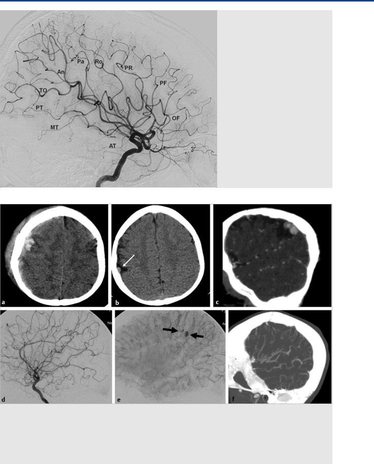
Fig. 16.4 The MCA branches can be best appreciated on the lateral view in this patient with a hypoplastic A1 segment. OF, orbitofrontal; PF, prefrontal; PR, pre-Rolandic; Ro, Rolandic; Pa, parietal; An, angular; TO, temporooccipital, PT, posterior temporal; MT, middle temporal; AT, anterior temporal.
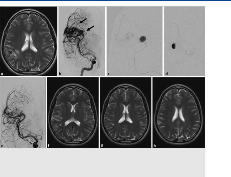
Fig. 16.5 A 15-month-old girl suffered a fall with skull fracture, a small subdural hematoma, and a contusional brain hemorrhage (unenhanced CT in a). The patient made an excellent recovery with no residual motor deficit. On follow-up imaging 3 months later, an encephalomalacic defect is seen. In the region of the previous fracture, a small extra-axial soft tissue density is seen (arrow in b), prompting further evaluation with CTA and, subsequently, digital subtraction angiography. CTA (c), sagittal reconstruction through the area of interest, demonstrates two adjacent aneurysmal outpouchings that were confirmed by digital subtraction angiography to originate from the distal Rolandic artery (right ICA injection lateral view in arterial [d] and capillary [e] phases). Contrast stagnation was seen in the aneurysms in the capillary phase (arrows in e). Given the eloquence of the affected vessel and the beginning thrombosis of the aneurysm, conservative management was selected. A follow-up CTA (f) demonstrated complete obliteration of the aneurysm.
78
The Cortical Branches of the Middle Cerebral Artery
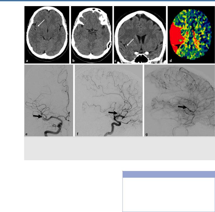
Fig. 16.6 Unenhanced CT in a 56-year-old female patient who presented with left-sided neglect, mild left upper limb weakness, and dysarthria demonstrates a hyperdense “dot” sign (arrows in a,c) in the right sylvian fissure as a sign of a distal MCA branch (M2) occlusion that was confirmed by CTA (not shown) and CT perfusion (TTP Map; d). Angiography of the right ICA in anteroposterior (e) and lateral (f,g) views demonstrates the sudden cutoff of the dominant inferior division of the right MCA trifurcation (arrows) with insufficient collateral flow to the distal MCA territory (belated phase on the lateral view injection). Case continues in Fig. 16.7.
the cortical territory they supply, which will lead to the following MCA “branches”: orbitofrontal, prefrontal, pre-Rolandic, Rolandic, parietal, angular, temporooccipital, posterior temporal, and middle and anterior temporal. These branches are best appreciated on the lateral view ( Fig. 16.4).
16.3 Clinical Impact, Additional
Information and Cases
See Fig. 16.5, Fig. 16.6, Fig. 16.7, Fig. 16.8, Fig. 16.9, and Fig. 16.10.
Pearls and Pitfalls
●Cortical branches of the MCA are named depending on the cortical territory they supply and are therefore best evaluated from distal to proximal on the lateral view.
●The MCA bifurcates into an inferior and a superior division that are in hemodynamic equilibrium with each other and that supply, respectively, the temporal and the frontal lobes and share the supply to the parietal lobe.
79
The Cortical Branches of the Middle Cerebral Artery
Fig. 16.7 A stent retriever was placed in the dominant inferior trunk, as verified by the angiographic run during retriever expansion (a; the small arrow points to the distal end). Follow-up angiography after a single pass demonstrated complete reopening of the vessel (b,c). On follow-up MR (axial diffusion-weighted imaging; d–f), a small area of (clinically silent) ischemia and persistent opening of the MCA branches on MR angiography (g) is seen.
Fig. 16.8 A patient with endocarditis and vegetations on his mitral valve presented with a right occipital hemorrhage (MR imaging with T1 [a] and susceptibility weighting [b]) and embolic hits (diffusion-weighted imaging in c). CTA in axial (d) and sagittal (e) cuts demonstrates a distal aneurysm as the source of the hemorrhage (arrows). As the patient was scheduled for urgent cardiac valve replacement surgery, embolization of the presumed mycotic aneurysm was requested. Right ICA injection in lateral view before embolization (f) demonstrated the peripheral MCA aneurysm that arises from the temporooccipital branch of the MCA that was subsequently catheterized. The aneurysm was occluded with glue embolization (immediate postembolization result [g]).
80
The Cortical Branches of the Middle Cerebral Artery
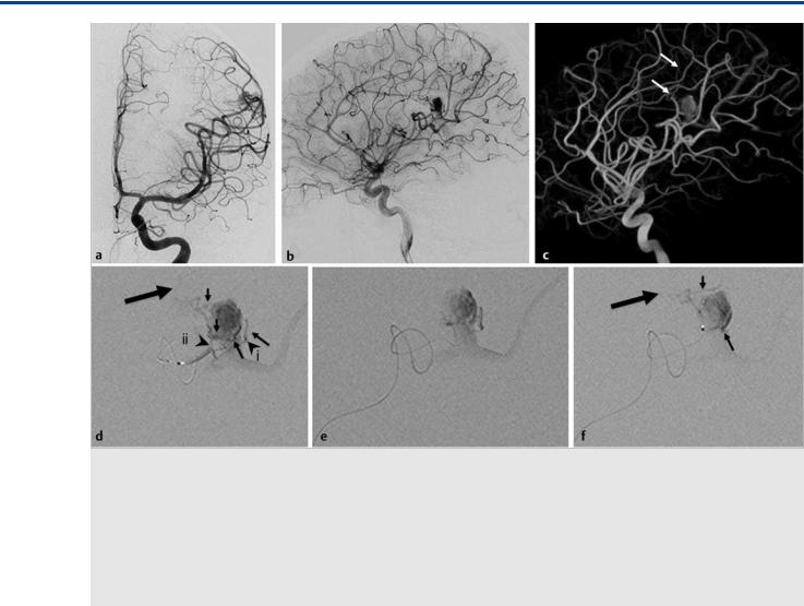
Fig. 16.9 A 14-year-old boy presented with seizures. On investigation, a brain arteriovenous malformation (AVM) was found that was subsequently evaluated by conventional angiography for potential embolization. Left ICA injections in anteroposterior (a) and lateral view (b) and 3D rotational angiography in a semioblique view (c) demonstrate that the AVM was fed by the Rolandic artery and that, distal to the AVM nidus, the feeding vessel to the precentral gyrus continued (arrows in c). Microcatheter injections into the proximal Rolandic artery (d) demonstrate four feeding arteries (small black arrows) into the shunt. The Rolandic artery continues distally (large arrow) to feed the primary motor cortex. Subsequently, injections were performed at the positions denoted by the arrowheads (i and ii). (e) The microcatheter injection in position (i). This is a safe vessel to embolize, as it is of the terminal type, with a sufficient security margin to the feeding artery. However, glue will likely enter the venous pouch and occlude the vein, which is the only venous outlet of the AVM. If additional arterial input is still present, bleeding of the AVM is likely to happen. Microcatheter injection in position (ii) (f) demonstrates two small vessels entering the AVM (arrows) that arise “en passage” from the Rolandic artery. The faint distal continuation to the ICA is again seen. Given these anatomical and angio-architectonic considerations, no embolization was performed.
81
The Cortical Branches of the Middle Cerebral Artery
Fig. 16.10 MR imaging in T2 weighting (a) demonstrates a right parietal AVM in a young patient with unspecific symptoms. There is peripheral edema surrounding a focal deep-seated outpouching of the AVM. Right ICA injection (b) demonstrates an enlarged leptomeningeal penetrating artery that feeds the medial (deep) portions of the AVM in which MR had demonstrated the outpouching (arrow in b). This feeder was subsequently catheterized and revealed a large intranidal aneurysm (c,d) that was subsequently embolized (immediate postembolization result seen in e). The patient underwent subsequent gamma knife radiosurgery, and 2-year follow-up axial T2 W MR sequences (f,g,h), show that the AVM was obliterated in a neurologically intact individual.
Further Reading
[1]Gibo H, Carver CC, Rhoton AL, Jr, Lenkey C, Mitchell RJ. Microsurgical anatomy of the middle cerebral artery. J Neurosurg 1981; 54: 151–169
[2]Morris P. Practical Neuroangiography. 3rd ed. Philadelphia: Lippincott, Williams & Wilkins; 2013
[3]Umansky F, Gomes FB, Dujovny M et al. The perforating branches of the middle cerebral artery. A microanatomical study. J Neurosurg 1985; 62: 261–268
[4]van der Zwan A, Hillen B, Tulleken CAF, Dujovny M, Dragovic L. Variability of the territories of the major cerebral arteries. J Neurosurg 1992; 77: 927–940
[5]van der Zwan A, Hillen B, Tulleken CAF, Dujovny M. A quantitative investigation of the variability of the major cerebral arterial territories. Stroke 1993; 24: 1951–1959
82
The Leptomeningeal Anastomoses
17 The Leptomeningeal Anastomoses
17.1 Case Description
17.1.1 Clinical Presentation
A 30-year-old man with history of prosthetic mitral valve replacement associated with rheumatic disease was poorly compliant with his anticoagulation therapy regimen. He presented to the emergency department with aphasia and left-sided hemiplegia, with the arm more pronounced then the leg. Because he had an absolute contraindication to the use of intravenous recombinant tissue plasminogen activator, he was brought to the angio-suite for mechanical thrombectomy.
17.1.2 Radiologic Studies
See Fig. 17.1, Fig. 17.2.
17.1.3 Diagnosis
Acute stroke resulting from embolic occlusion of the superior division of the middle cerebral artery (MCA) with excellent leptomeningeal collateral circulation.
17.2 Anatomy
Leptomeningeal anastomoses (LMAs), or leptomeningeal collaterals, are small arteriolar connections (~50–400 µm) between two cerebral arteries supplying two di erent but adjacent cortical territories. They were initially described by Sir Thomas Willis in 1684, but the first well-documented work demonstrating their presence was done by Heubner in 1874. These vessels form an extensive network that provides a potential route for collateral perfusion, together with the large arterial communications and the circle of Willis.
In LMAs, blood can flow in both directions, depending on the hemodynamic and metabolic needs, thus allowing retrograde perfusion of adjacent territories. Their presence, number, size, and location have a high degree of variability between individuals as well as between hemispheres in the same person. They tend to be more extensive along the convexity, connecting the MCA with the anterior cerebral artery (ACA) and posterior cerebral artery (PCA) territories, but there are also LMAs connecting the ACA and PCA territories (precuneus and splenium of the corpus callosum), as well as both ACA territories (through callosal arteries) and between distal branches of the MCA, PCA, and ACA ( Table 17.1).
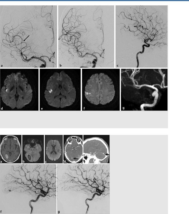
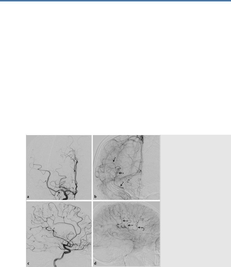
Fig. 17.1 Right internal carotid artery (ICA) angiogram in anteroposterior (AP) (a,b) and lateral (c,d) views in arterial (a,c) and late capillary (b,d) phase. There is occlusion of the superior division of the middle cerebral artery (MCA), with a focal extrinsic filling defect in the M1 trunk (arrow). The late-capillary-phase angiograms show retrograde filling of the superior division of the MCA via LMAs from the ACA (thin arrows in b,d). Case continued in Fig. 17.2.
83
The Leptomeningeal Anastomoses
Fig. 17.2 Right ICA angiogram in AP view after thrombectomy (a) demonstrates complete recanalization of the superior division of the MCA with good antegrade flow and no distal filling defects. Control unenhanced axial computed tomography 24 hours after thrombectomy (b,c) shows an infarct in the right striatum but no cortical infarct in the territory of the superior division of the MCA, as the cortex received collateral supply during MCA trunk occlusion, whereas the perforators did not.
Table 17.1 Territories, arteries involved, and location of the most common leptomeningeal anastomoses: Orbito-frontal (MCA) with fronto orbital and fronto-polar (ACA).
Territory |
Anastomosing Arteries |
Location of the Anastomosis |
|
|
|
MCA and ACA |
Orbito-frontal (MCA) with fronto-orbital and fronto-polar (ACA) |
Inferior and middle frontal gyrus |
|
|
|
|
Pre-frontal artery (MCA) with anterior/middle internal frontal artery (ACA) |
Superior frontal sulcus |
|
|
|
|
Pre-central artery (MCA) with posterior internal frontal artery (ACA) |
Pre-central sulcus |
|
Central artery (MCA) with paracentral artery (ACA) |
Central sulcus |
|
|
|
|
Parietal artery (MCA) with superior parietal (ACA) |
Post-central sulcus or intraparietal sulcus |
|
|
|
MCA and PCA |
Angular or posterior temporal artery (MCA) with parieto-occipital artery (PCA) |
Parieto-occipital or intraparietal sulcus |
|
|
|
|
Temporal arteries (MCA) with inferior temporal trunk branches (PCA) |
Middle temporal sulcus |
ACA and PCA |
Superior parietal or pericallosal arteries (ACA) with parieto-occipital artery (PCA) |
Parieto-occipital sulcus |
|
|
|
|
Pericallosal artery (ACA) with splenial artery (PCA) |
Splenium of the corpus callosum |
|
|
|
ACA = anterior cerebral artery; MCA = middle cerebral artery; PCA = posterior cerebral artery
The ability of LMAs to compensate for a vascular occlusion depends on four main factors: presence and number of LMAs, systemic blood pressure, onset of the occlusion, and age of the patient. As stated earlier, the number and diameter of the LMAs vary significantly among di erent individuals, but the pressure gradient magnitude between the una ected and a ected vascular territory, and thus the flow rate, is influenced by the systemic blood pressure. Nevertheless, it has been seen that patients with chronic systolic hypertension will have less-func- tional LMAs because of impairment of the cerebral blood flow autoregulation. Patients with a gradual onset of a vascular occlusion (moyamoya syndrome or intracranial or extracranial stenosis) develop a more extensive LMA network that allows for better compensatory collateral flow changes than does an abrupt arterial occlusion. The functional compensatory capacity of LMAs also diminishes with age. This is probably mediated by
a decrease in the number of LMAs, as well as “sti ening” of these vessels mediated by atherosclerosis, decreasing their ability to dilate properly in response to flow or metabolic demands.
17.3 Clinical Impact
At this time, there is a general consensus that the ability of LMAs to compensate for a vascular occlusion is one of the main determinants of outcome in patients with acute stroke and is, therefore, critical in patient selection for intra-arterial endovascular stroke treatments. Good collateral flow via the LMAs is assumed to be independently associated with a favorable outcome because of the capacity to maintain the ischemic penumbra until reperfusion occurs. Computed tomographic angiography gives a rough estimation of the collateral flow, specifically
84
The Leptomeningeal Anastomoses
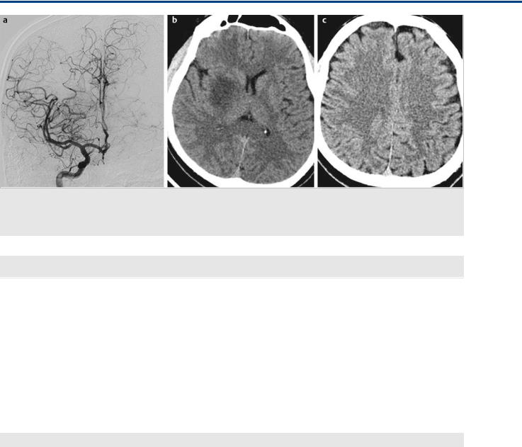
Fig. 17.3 A 66-year-old man presenting with an acute subarachnoid hemorrhage. Right vertebral artery angiogram in lateral view before (a) and after
(b) coil occlusion of the distal P1 segment for treatment of a right PCA dissecting aneurysm located in the P1/P2 segment junction. Right ICA angiogram in lateral view immediately after the procedure (c) shows retrograde filling of the distal PCA branches via LMAs, with contrast filling seen up to the P3 segment of the PCA.
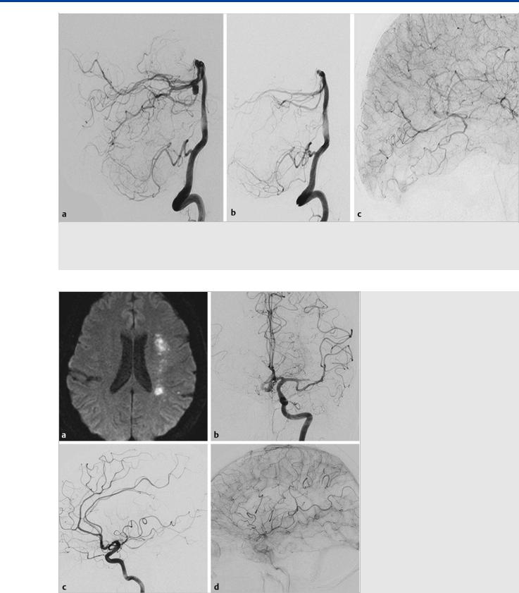
Fig. 17.4 A 57-year-old woman with acute onset of focal neurological deficits. Diffusion-weighted MRI (a) shows acute ischemic lesions in the ACA/ MCA watershed territory. Left ICA angiogram in AP view (b) shows an occlusion in the superior division of the left MCA. Left ICA angiogram in lateral view in arterial (c) and late (d) capillary phase shows significantly decreased antegrade flow through the superior division of the MCA, with retrograde filling of the distal cortical branches through LMAs from the ACA.
85
The Leptomeningeal Anastomoses
Fig. 17.5 LMAs are often recruited in high-flow AVMs because of the “sump” effect. This can be explained by the low resistance of the high-flow shunt, as there is no intervening capillary bed at the level of the shunt, with subsequently increased flow from neighboring territories. The high-flow nature of the AVM may also lead to relative perinidal hypoxemia with secondarily enlarged leptomeningeal collaterals. Both factors will lead to a “shift of the watershed” (i.e., an apparent supply from neighboring branches to the AVM) as indicated in this example. Right (a) and left (b) ICA angiograms in AP view demonstrate that the ACA branches do not feed the AVM proper but are only secondarily recruited to anastomose via the leptomeningeal vessels to the MCA branches that supply the AVM. Caution must be taken not to include the secondarily recruited vessels in the treatment plan.
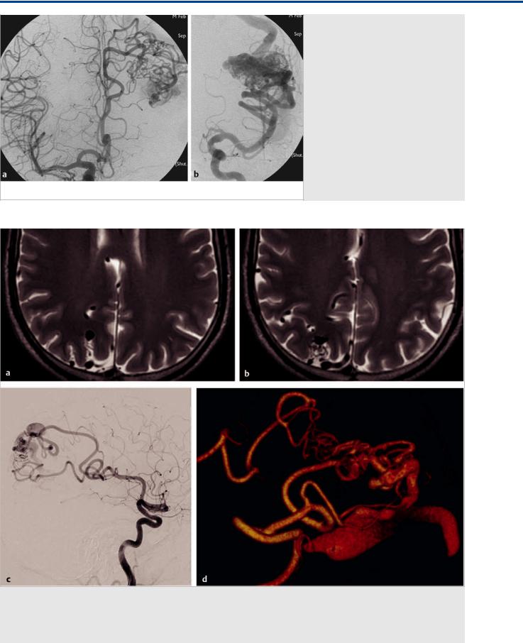
Fig. 17.6 Although in its most dramatic form the shift of the watershed resulting from leptomeningeal collaterals is visible from one large vessel territory into the other (ACA to MCA or vice versa), it may also be present between neighboring distal pial branches, as visualized in this case with a fistulous high parietal AVM, where high-resolution T2-weighted MRI (a,b) demonstrate dilated transgyral vessels and right ICA angiograms in lateral view (c) and 3D rotational reconstruction (d) demonstrates that the angular artery (via dilated leptomeningeal branches) secondarily contributes to the supply of the posterior parietal branch that is the sole feeding vessel of the AVM.
86