

Книги по МРТ КТ на английском языке / Neurovascular anatomy in interventional neuroradiology Krings et al 2015
.pdfThe Superficial Cortical Veins
be absent. In this case, the anterior component will drain toward the sphenoid ridge sinuses and the posterior segment to the vein of Trolard, the vein of Labbé, or both.
The classic notion that the SMV drains into the so-called sphenoparietal sinus has recently been challenged. The sphenoparietal sinus actually corresponds to a combination of two separate venous structures: the parietal portion of the anterior branch of the middle meningeal veins and a dural channel located under the lesser sphenoid wing, called the sinus of the lesser sphenoid wing or sphenobasilar sinus wing. It was shown that the SMV never connected to the anterior branch of the middle meningeal veins or the sinus of the lesser sphenoid wing, although it might drain into the cavernous, paracavernous, and/or laterocavernous sinuses. This so-called cavernous capture of the SMV is not seen in the embryo or during early infancy and occurs during the first 6 to 12 months of age.
The three aforementioned venous groups with their respective anastomotic collector veins exist in a hemodynamic balance in a reciprocal relationship. The anatomic configuration of these veins can range from a balanced network of three separate or interconnected anastomotic draining veins to a dominant anastomotic vein, with the two others being hypoplastic. The vein of Trolard or Labbé may be completely absent, but the SMV, at least in part, will almost invariably be present.
34.3 Clinical Impact
The main complication of not recognizing a single dominant system is a venous infarct after surgery or endovascular procedures if the drainage route is inadvertently compromised. For example, in pterional craniotomies for clipping of middle cerebral artery aneurysms, patients with a dominant SMV are at a higher risk of venous infarct if the vein is occluded during surgery. In patients with a transvenous approach for dural arteriovenous fistulas of the transverse sinus, the opening of a dominant vein of Labbé should be preserved while occluding the sinus. If a transarterial approach is used, the operator should be careful to avoid “overshooting” the fistula (i.e., pushing too much liquid embolic material too deep into a dominant Trolard or Labbé vein).
34.4 Additional Information and
Cases
When a cortical vein does not fill in the venous phase of a cerebral angiogram, the question is whether that vein is congenitally absent or is present, but filled with thrombus. Clinical presentation and ancillary findings in the noninvasive imaging studies are crucial to answering this question properly. From an angiographic point of view, it helps to assess the individual venous anatomy characteristics. If there is a clearly dominant single collector or two large collectors that are patent, the absent vein is probably hypoplastic or absent. If, in contrast, other patent venous collectors are small, the absent vein is more likely to be thrombosed. The exception would be the SMV, which, at least in part, is almost always present, so complete absence of the SMV in the venous phase is likely to be a venous thrombosis/occlusion ( Fig. 34.2; Fig. 34.3; Fig. 34.4; Fig. 34.5;Fig. 34.6).
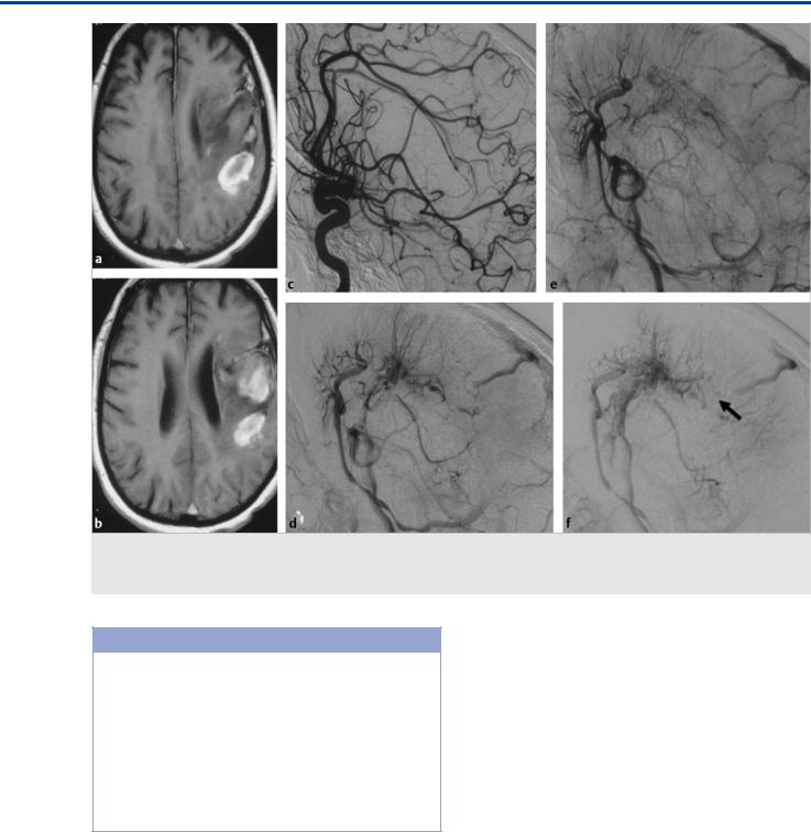
Fig. 34.6 ICA injection lateral view in a patient with a brain AVM. The AVM is located in the tributary territory to both the Labbé vein and the middle superficial cerebral vein, and there is subsequent drainage into both. Because of venous obstruction involving the outlets of both major drainage routes, there is secondary rerouting into cranially directed veins, leading to widespread interference with normal venous drainage. This type of pattern has been associated to presentation with seizures resulting from venous congestion.
Pearls and Pitfalls
●There are three main venous collectors for the cortical draining veins: the vein of Trolard, the vein of Labbé, and the SMV. These three collectors exist in a hemodynamic balance.
●If there is one single or two dominant collectors, the third one may be absent, with an exception made for the superficial middle cerebral vein (SMV), of which at least part will almost invariably be present.
●Nonvisualization of the vein of Labbé or Trolard during the venous phase may be normal, but if the SMV is not demonstrated, venous thrombosis or occlusion is likely.
●The occlusion of a dominant collector will almost invariably result in a venous infarct because of venous congestion, as there is no good collateral venous drainage. As such, occluding a large collector during surgery or endovascular procedures has to be avoided.
●The individual variability between size, configuration, and location of these collectors explains the high variability in the patterns of venous ischemia.
●Widespread venous congestion or a long pial course of the cortical draining vein has been implicated in clinical presentation with seizures in patients with brain AVMs.
177

The Superficial Cortical Veins
Further Reading
[1]Lasjaunias P, Berenstein A, ter Brugge KG. Surgical Neuroangiography. Vol. 1. 2nd ed. Berlin: Springer; 2006
[2]Rhoton AL, Jr. The cerebral veins. Neurosurgery 2002; 51 Suppl: S159–S205
[3]San Millán Ruíz D, Fasel JH, Rüfenacht DA, Gailloud P. The sphenoparietal sinus of Breschet: does it exist? An anatomic study. AJNR Am J Neuroradiol 2004; 25: 112–120
[4]Scott JN, Farb RI. Imaging and anatomy of the normal intracranial venous system. Neuroimaging Clin N Am 2003; 13: 1–12
[5]Shankar JJ, Menezes RJ, Pohlmann-Eden B, Wallace C, ter Brugge K, Krings T. Angioarchitecture of brain AVM determines the presentation with seizures: proposed scoring system. AJNR Am J Neuroradiol 2013; 34: 1028–1034
178
The Transmedullary Veins
35 The Transmedullary Veins
35.1 Case Description
35.1.1 Clinical Presentation
A 61-year-old previously healthy man came to medical attention because of mild cognitive deficits and slight subjective gait instability.
35.1.2 Radiological Studies
See Fig. 35.1, Fig. 35.2, and Fig. 35.3.
35.1.3 Diagnosis
Choroidal arteriovenous malformation (AVM) with reflux into transmedullary veins leading to hydrocephalus.
35.2 Embryology and Anatomy
The transcerebral veins are also called medullary or anastomotic veins, as they interconnect the superficial with the deep
venous systems. Embryologically, these veins become apparent in the 40-mm embryo as a plexus of fine, straight veins that pass from the ependymal layer to the surface of the cortex, where they drain into pial collectors. There are two transcerebral venous systems present that may interconnect with each other via deep-seated venular anastomoses: the superficial and the deep medullary veins. While the former drain the white matter toward the surface from a depth of 1–2 cm, the latter drain the remainder of the white matter toward the ventricle centripetally into the subependymal veins of the lateral sinuses. Direct anastomotic veins between the superficial cortical and deep subependymal systems may be present.
The deep veins of the frontal and parietal lobes have a fanshaped organization and converge at the superolateral angle of the lateral ventricle, with frontal veins joining the septal and anterior caudate veins and the parietal veins draining toward the thalamostriate veins. The occipital lobe veins join the lateral atrial veins, whereas the temporal lobe transmedullary veins take an ascending direction to join the inferior ventricular veins.
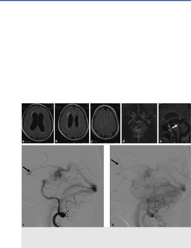
Fig. 35.1 Axial fluid-attenuated inversion recovery (FLAIR) weighted scans in multiple cuts (a,b,c) demonstrate mild hydrocephalus with increased size of the ventricles, decreased width of the sulci, and periventricular FLAIR hypersignal as a sign for decreased transependymal CSF reabsorption. The time-of-flight MRA (d) shows a choroidal AVM, and the midsagittal T1-weighted scan (e) proves patency of the aqueduct (arrow). No other cause for the patient’s hydrocephalus was identified. Conventional angiography (vertebral artery injection, lateral views on early and late phases f,g) revealed the AVM, which drained not only toward the straight sinus but also into the transmedullary veins (arrows in f,g). Case continued in Fig. 35.2.
179
The Transmedullary Veins
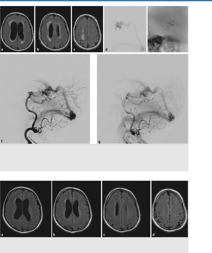
Fig. 35.2 Within the next 5 months, the patient’s symptoms became worse, with significant impairment of cognitive function and corresponding worsening of his hydrocephalus (axial FLAIR weighted scans, a,b,c), with ventricular widening and increased interstitial edema suggesting further decreased transependymal CSF reabsorption. There was no evidence for obstruction of the ventricular system, and it was proposed that the hyperpressure in his transependymal veins after arterialization was related to an impaired CSF reabsorption through the transmedullary veins. After partial embolization ([d] microcatheter position in the posterior lateral choroidal artery; [e] glue cast) that led to obliteration of the transmedullary drainage (conventional angiography postembolization, lateral view [f,g]), the symptoms reversed. Case is continued in Fig. 35.3.
Fig. 35.3 MR follow-up 3 weeks after the embolization demonstrated marked improvement of the transependymal CSF reabsorption on various axial FLAIR sections (a–d). The hydrocephalus had decreased, indicating it was indeed the arterialization of the transmedullary veins that could be held responsible for the decreased CSF reabsorption.
180
The Transmedullary Veins
Angiographically, these vessels are typically not seen; however, they will be visualized in the setting of arterialization of the veins (because of an arteriovenous shunt), significant outflow restrictions, or in the presence of developmental venous anomalies (DVAs). It was Pierre Lasjaunias who pointed out that these DVAs are, in fact, extreme variations of the normal trans-
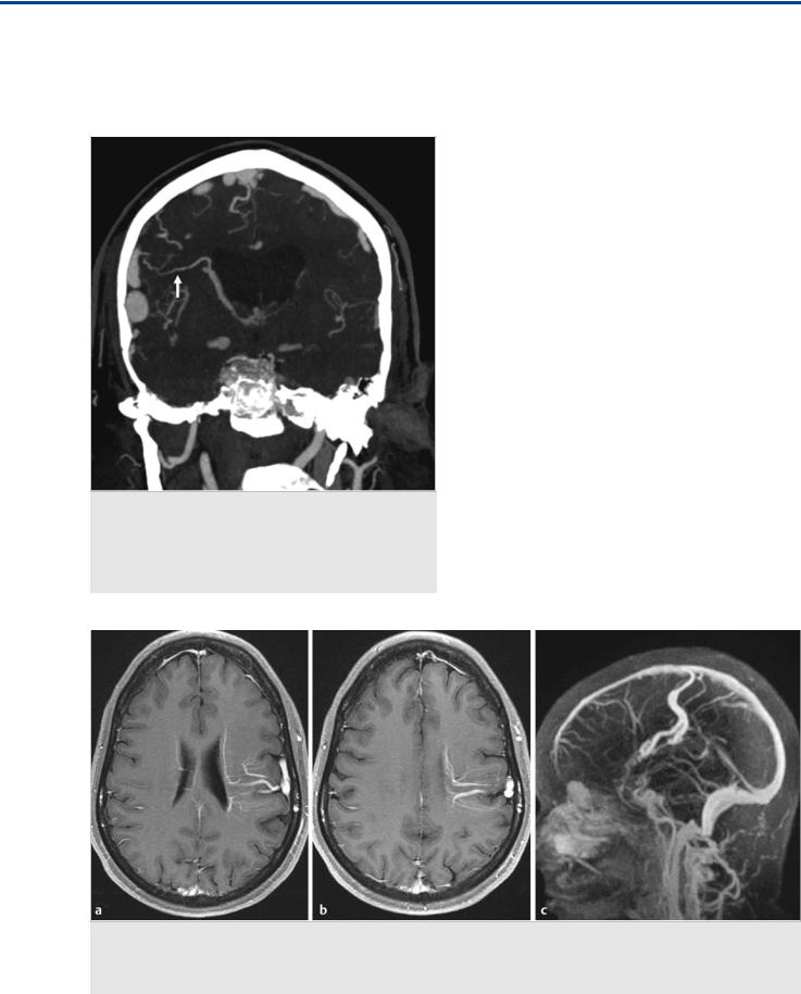
Fig. 35.4 In a patient with a high-flow pial arteriovenous fistula with multiple venous pouches, drainage is directed in part into the deep venous system (note the different caliber of the thalamostriate veins on both hemispheres) through a dilated transmedullary vein (arrow). If in these scenarios hydrocephalus is present, it is believed to be related to decreased CSF reabsorption via the transmedullary veins.
medullary veins that are necessary for the drainage of white and gray matter. They consist of converging dilated medullary veins that drain centripetally and radially into a transcerebral collector that opens into either the superficial subcortical or deep pial veins. They serve as normal drainage routes of the brain tissue, as the habitual venous drainage of their territory is absent. Their etiology and mechanism of development are unknown, but it is currently accepted that they act as a compensatory system of cerebral parenchymal venous drainage because of an early failure, abnormal development, or intrauterine occlusion of normal capillaries or small transcerebral veins.
35.3 Clinical Impact
The aforementioned considerations, as well as the described clinical case, indicate that transmedullary veins may play a role in two di erent types of clinical scenarios: disorders of reabsorption of cerebrospinal fluid (CSF) and in patients with DVAs. Concerning the former, CSF is absorbed not only via arachnoid granulations (pacchionian bodies), as in the widely accepted bulk flow theory, but also via physiological transependymal flow at the level of the brain capillaries. Increased venous pressures in the periventricular and transmedullary veins, as a consequence of arteriovenous shunting toward periventricular and transmedullary veins, as seen in the index patient, will therefore impede e cient CSF absorption by reducing the pressure gradient between the capillary bed and surrounding CSF spaces. Drainage into the transmedullary veins is, however, rare and is typically seen only if deep or superficial venous systems are overloaded (because of downstream narrowing or very high flow) or if normal drainage pathways are occluded or nonfunctional (e.g., in babies with vein of Galen AVMs). In these instances, the collateral transcerebral anastomotic outlets will have to be recruited. In patients with vein of Galen AVMs, the oftenvisualized hydrocephalus is therefore related to the same
Fig. 35.5 MRI with contrast-enhanced axial T1-weighted images (a,b) reveals tubular enhancing structures within the periventricular and cortical regions of the left frontoparietal lobe with a caput medusae-like appearance. Contrast-enhanced MR venography (c) in sagittal view confirms the presence of a deepto superficial-type DVA, draining into the superior sagittal sinus. Note the missing connection of the deep periventricular veins on the left to the ventricular atrial and thalamostriate veins, which are present on the unaffected right side.
181
The Transmedullary Veins
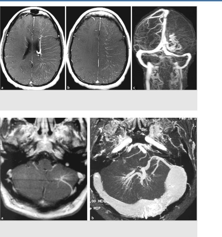
Fig. 35.6 MRI with contrast-enhanced axial T1-weighted images (a,b) reveal tubular enhancing structures within the periventricular and cortical regions of the left cerebral hemisphere, with a collecting vein at the left lateral ventricle. MR venography (c) in coronal view confirms the presence of a superficialto deep-type DVA, draining into the deep venous system. Note the paucity of the superficial cortical venous system on the left compared with on the right.
Fig. 35.7 Contrast-enhanced axial T1-weighted images in two different patients (a,b) reveal posterior fossa DVAs. The larger lesion in the second patient (b) is also associated with a dural sinus malformation. The larger the lesion, and the bigger the territory affected by the misbalanced venous drainage, the higher the likelihood for these lesions to become symptomatic.
pathomechanical concept as in our index patient, which explains why extraventricular drainage will not ameliorate the hydrocephalus in these vascular shunts.
Concerning the clinical significance of transcerebral veins in DVAs, it has to be first pointed out that DVAs are benign anatomical variations and are, therefore, usually incidentally discovered. It is only rarely that they become symptomatic. As DVAs are
extreme variations of normal venous drainage, a single collector drains an abnormally large parenchymal territory. This can lead to a more fragile venous outflow system, as the single venous collector can be overloaded, accounting for the dilated medullary veins. Therefore, AVMs employing a DVA as their venous outflow are more likely to rupture as compared to AVMS that can drain into both the deep and the superficial venous systems.
182
The Transmedullary Veins
Fig. 35.8 T1-weighted scans (a,b) in this patient with acute onset of hemiparesis and headache demonstrate a left frontoparietal hemorrhage. On conventional angiography (c–f, lateral view of left internal carotid artery injection at arterial and capillary early and late venous phases), a complex DVA is seen in with acute thrombosis in a posteriorly located collecting vein (arrow in f).
Pearls and Pitfalls
●Transmedullary veins connect the deep and superficial venous systems.
●They are usually “dormant” channels that may be recruited in postnatal life if an arteriovenous shunt has overloaded either the deep or the superficial system because of high flow or associated outflow obstructions.
●In patients with DVAs, these anastomoses are no longer present, as either the deep system drains superficially or vice versa. Patients with a DVA, therefore, have less compensatory capabilities of their venous system.
Further Reading
[1]Ebinu JO, Matouk CC, Wallace MC, Terbrugge KG, Krings T. Hydrocephalus secondary to hydrodynamic disequilibrium in an adult patient with a choroi- dal-type arteriovenous malformation. Interv Neuroradiol 2011; 17: 212–216
[2]Geibprasert S, Pereira V, Krings T, Jiarakongmun P, Lasjaunias P, Pongpech S. Hydrocephalus in unruptured brain arteriovenous malformations: pathomechanical considerations, therapeutic implications, and clinical course. J Neurosurg 2009; 110: 500–507
[3]Jimenez JL, Lasjaunias P, Terbrugge K, Flodmark O, Rodesch G. The trans-cere- bral veins: normal and non-pathologic angiographic aspects. Surg Radiol Anat 1989; 11: 63–72
[4]Lasjaunias P, Burrows P, Planet C. Developmental venous anomalies (DVA): the so-called venous angioma. Neurosurg Rev 1986; 9: 233–242
[5]Pereira VM, Geibprasert S, Krings T et al. Pathomechanisms of symptomatic developmental venous anomalies. Stroke 2008; 39: 3201–3215
35.4 Additional Information and
Cases
See Fig. 35.4, Fig. 35.5, Fig. 35.6, Fig. 35.7, andFig. 35.8.
183
The Deep Venous System I: Internal Cerebral Veins, Tributaries, and Drainage
36 The Deep Venous System I: Internal Cerebral Veins, Tributaries, and Drainage
36.1 Case Description
36.1.1 Clinical Presentation
A 64-year-old woman presented with seizures and progressive headaches. Outside computed tomography revealed a large mass centered at the tentorial incisura, and she presented to neurosurgery.
36.1.2 Radiologic Studies
See Fig. 36.1, Fig. 36.2, and Fig. 36.3.
36.1.3 Diagnosis
Tentorial incisura meningioma with occlusion of the straight sinus and rerouting of the deep venous drainage.
36.2 Embryology and Anatomy
The deep venous system consists of the internal cerebral veins (ICVs) and their tributaries and, morphologically speaking, the basal vein of Rosenthal system, which is discussed in Case 37. During embryogenesis, the first vein to drain the developing brain is the median prosencephalic vein (MPV; or vein of Markowski), a single, temporary midline vein that drains the
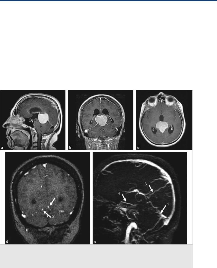
Fig. 36.1 In this patient with a tentorial incisura meningioma (a–c; T1-weighted postcontrast MRI), the major question to be answered before surgical removal is whether the straight sinus is functional and, if not, how the deep brain drains, as this will determine which veins to spare during surgery and which approach to take. Coronal MR venography (d) with sagittal reconstructions (e) suggests occlusion of the vein of Galen with a straight sinus stump (1), persistence of a left-sided tentorial sinus (2), superior drainage into a medial parietal vein (3), and anterior drainage of the right basal vein of Rosenthal (4). Digital subtraction angiography was performed to further elucidate these findings. Case continued in Fig. 36.2.
184
The Deep Venous System I: Internal Cerebral Veins, Tributaries, and Drainage
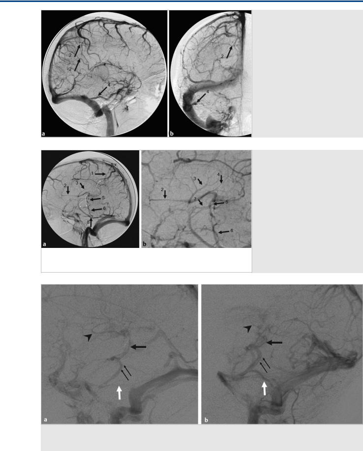
Fig. 36.2 Right internal carotid artery injection lateral (a) and AP (b) in the venous phase demonstrates drainage of the ICVs, rather than in the vein of Galen, craniomedially into two medial parietal veins (2) toward the superior sagittal sinus. The basal vein of Rosenthal is seen to drain laterally via the inferior temporal vein (4) toward the transverse sinus. The small white arrows denote the collateral pathways. Case continued inFig. 36.3.
Fig. 36.3 Left internal carotid artery injection in the venous phase in lateral view (a, enlarged view in b) demonstrates drainage to the medial parietal veins (1) toward the superior sagittal sinus, but also drainage from the septal veins (2), the thalamostriate veins (3), and the posterior atrial veins (4) into the ICVs, and from there, via an inferior thalamic vein (5) to the lateral mesencephalic vein (6), which opens to the superior petrosal vein. This “epsilon” shape of the venous drainage is typically seen in vein of Galen AVMs, where the deep venous system has not gained access to a normally developed straight sinus drainage. Additional drainage of the basal vein of Rosenthal (7) in the present case is through the inferior temporal vein (8). The small curved white arrows denote the collateral pathways.
Fig. 36.4 The epsilon-shaped drainage of the deep venous system in two different patients (a,b). The thalamostriate veins (arrowhead) drain via the inferior thalamic vein (black arrow) toward the lateral mesencephalic vein (small black arrows), which drains via the superior petrosal sinus (white arrow) into the sigmoid sinus. Note the absence of the straight sinus in (b).
185
The Deep Venous System I: Internal Cerebral Veins, Tributaries, and Drainage
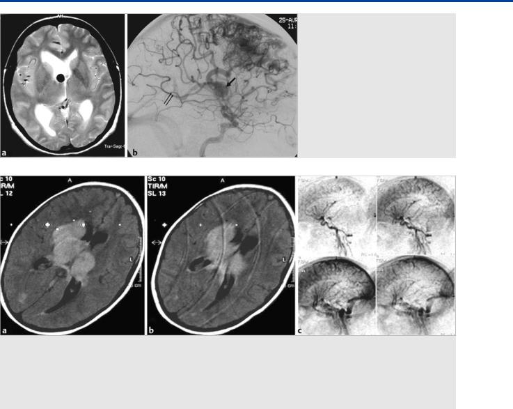
Fig. 36.5 Axial T2-weighted MRI (a) and lateral view of a right internal carotid artery injection (b). Hydrocephalus in a patient with an unruptured AVM due to occlusion of the foramen of Monro by a venous pouch related to a focally dilated anterior thalamostriate vein that served as drainage route of a right frontal anterior cerebral artery–fed AVM. Note the deep venous drainage of this AVM, as indicated by early opacification of the vein of Galen (double arrow).
Fig. 36.6 Axial FLAIR weighted sequences (a,b) in a 7-year-old girl with progressive loss of consciousness over the last few hours and a newly developed anisocoria demonstrates edematous changes bilaterally in the thalamus and in the right caudate. Contrast-enhanced, time-resolved MRA on sagittal cuts demonstrates the absence of the straight sinus, the ICVs, and the basal vein of Rosenthal, indicating extensive internal cerebral venous thrombosis with significant venous congestion. The patient had complained about headaches over the course of the last 4 days and was severely dehydrated because of repeated vomiting. The thalamus is drained by multiple routes: its anterior portion can join the thalamostriate vein, the superior thalamic vein enters the ICV directly, and the posterior and inferior thalamic veins enter into the basal vein of Rosenthal or the lateral mesencephalic vein. The significant venous congestion seen in this child indicates widespread deep venous system thrombosis.
choroid plexus and the surrounding neural parenchyma. Progression of arterial supply and growth of neural structures will lead to formation of the paired ICVs and subsequent regression of the median prosencephalic veins as the ICV annexes the drainage of the choroid plexus. The most caudal part of the MPV persists as the vein of Galen. If an arteriovenous shunt has formed, however, between the choroidal arteries and the MPV prior to this annexation, the arterialization of the MPV will prevent its regression, and the MPV will persist as the major drainage of the so-called vein of Galen arteriovenous malformation (AVM), which is in fact a choroidal AVM that is draining through the embryonic precursor of the vein of Galen (i.e., the MPV). In these cases, the ICV will not connect to the deep venous system (i.e., the vein of Galen) and will have to find alternate drainage pathways.
In normal development, the ICV will drain most of the deep subependymal and choroid venous system. The ICV is formed at the foramen of Monro by the confluence of the septal and thalamostriate veins (the venous angle) as the most consistent and largest tributaries of the ICV. The septal veins drain the deep structures of the frontal lobe, the septum and the fornix. The
medial surface of the caudate head is drained by the anterior caudate vein, which opens into the thalamostriate vein, which receives additional supply from the transmedullary veins of the posterior frontal and anterior parietal lobes and the internal capsule. Despite its name, it does not play a major role in drainage of the thalamus. Along its posterior course within the tela choroidea, close to the midline, the ICV will receive the choroidal veins and additional subependymal inflow from the atrial veins and the superior thalamic vein, the vein of the corpus callosum, and the habenular veins. Both ICVs merge to form the vein of Galen between the pineal gland and the inferior aspect of the splenium. The vein of Galen is subarachnoid in location and will receive multiple other sources of venous drainage, acting as a true collecting vein with multiple potential venous outlets. It will connect bilaterally to the basal vein of Rosenthal (opening an anteromedial drainage route to the cavernous sinus and an anterolateral route to the temporal cortex via the inferior temporal vein). Superiorly, the vein of Galen will connect with the splenial and occipital veins, as well as the inferior sagittal sinus. Its major drainage route is through the straight sinus.
186