

- •Dedication
- •Preface
- •Acknowledgments
- •Figure Credits
- •Expert Consultants and Reviewers
- •Contents
- •Descriptive Terms for Normal Cells
- •Descriptive Terms for Abnormal Cells and Tissues
- •Epithelium
- •Glands
- •Introduction and Key Concepts for Connective Tissue
- •Cartilage
- •Bone
- •Introduction and Key Concepts for the Nervous System
- •Peripheral Blood Cells
- •Hemopoiesis
- •Introduction and Key Concepts for the Circulatory System
- •The Cardiovascular System
- •Introduction and Key Concepts for the Lymphoid System
- •Cells in the Lymphoid System
- •Introduction and Key Concepts for the Respiratory System
- •Conducting Portion
- •Respiratory Portion
- •Introduction and Key Concepts for the Urinary System
- •Introduction and Key Concepts for the Integumentary System
- •Oral Mucosa
- •Teeth
- •Introduction and Key Concepts for the Digestive Tract
- •Introduction and Key Concepts for the Endocrine System
- •Introduction and Key Concepts for the Male Reproductive System
- •Introduction and Key Concepts for the Female Reproductive System
- •Introduction and Key Concepts for the Eye
- •Introduction and Key Concepts for the Ear
- •Introduction
- •Preservation versus Fixation
- •Fixatives and Methods of Fixation
- •Sectioning and Mounting
- •Staining
- •Index

370 UNIT 3 ■ Organ Systems
Figure 19-13A |
Overview of the Placenta |
Figure 19-13B |
Fetal Portion of the Placenta |
Figure 19-14A |
Umbilical Cord |
Vagina |
|
Figure 19-14B |
Vagina |
Mammary Glands |
|
Figure 19-15A |
Overview of the Mammary Gland |
Figure 19-15B |
Inactive (Resting) Mammary Gland |
Figure 19-15C |
Active (During Pregnancy) Mammary Gland |
Figure 19-16A |
Nipple, Mammary Gland |
Figure 19-16B |
Clinical Correlation: Adenocarcinoma of the Breast (Breast Cancer) |
Synopsis 19-1 |
Clinical and Pathological Terms for the Female Reproductive System |
Introduction and Key Concepts for the Female Reproductive System
The female reproductive system comprises the ovaries, oviducts, uterus, vagina, external genitalia, and mammary glands. The external genitalia (vulva) includes the labia minora, labia majora, mons pubis, clitoris, and vestibule. Female secondary sex characteristics appear at puberty, along with the monthly menstrual cycle. This cycle of changes in the reproductive system is influenced by interactions among the hypothalamus, pituitary gland, ovaries, and uterus; related events occur periodically during each menstrual cycle (Fig. 19-8). The menstrual cycle is influenced by hormones including follicle-stimulating hormone (FSH), luteinizing hormone (LH), estrogen, and progesterone. These hormones cause changes in the female reproductive organs and their functions, promote development of follicles and oocytes, and produce an ideal environment for fertilization, implantation, and fetal growth. The female reproductive system plays an important role in the production and regulation of female hormones (estrogen and progesterone) and in the development and maintenance of female sex characteristics.
Ovaries
The ovaries are paired, almond-shaped structures located in the upper part of the pelvic cavity. Their size and position vary depending on the age and reproductive state of the individual. The ovaries are suspended by the mesovarium of the broad ligament and are attached to the uterus by the ligament of the ovary (Fig. 19-1). Each ovary has a cortex and medulla. The cortex contains numerous developing follicles in various stages as well as postovulatory structures, a corpus luteum, and several corpora albicans. Each developing follicle contains an oocyte. The medulla is composed of loose connective tissue and blood vessels, nerve fibers, and lymphatic vessels (Fig. 19-3A).
1.Primordial follicles: In the earliest stage of follicular development, primordial follicles rest at the periphery of the
cortex. Each primordial follicle consists of a primary oocyte surrounded by a single layer of squamous supporting cells called follicular cells (Fig. 19-4A,B). The oocyte is small (about 20–30 μm) and is in prophase (dictyotene) of meiosis I. The nucleus of the oocyte has a pale appearance and contains decondensed chromatin.
2.Primary follicles: At puberty, the primordial follicles begin to grow, the oocyte increases its size, and the supporting follicle cells also increase in size and become cuboidal cells. These follicle cells are now called granulosa cells. When the oocyte of the primary follicle is surrounded by a single layer of granulosa cells, the follicle is called a unilaminar primary follicle. As the oocyte increases in size, the granulosa cells build up more layers, and the follicle is called a multilaminar primary follicle (Fig. 19-5A,B). The zona pellucida, a gel-like layer between the oocyte and the granulosa cells, first appears in the multilaminar primary follicle (Fig. 19-5B).
3.Secondary follicles: As granulosa cells continue to proliferate, the follicle size increases, and spaces filled with follicular fluid (liquor folliculi) develop among the cells. These spaces merge to become a single large space called the antrum. The stromal cells that cover the follicle develop into a layer called the theca folliculi. The theca folliculi is well developed in the secondary follicle, and it includes the theca interna and theca externa (Fig. 19-6A).
4.Graafian (preovulatory) follicle: In its final stage, the follicle reaches a maximum size of up to 25 mm (2.5 cm). This follicle has a large antrum filled with liquor folliculi. It has reached its mature stage and is ready to release the oocyte (ovulation). The oocyte has reached its maximum size, and is embedded in a mound of granulosa cells that protrude into the antrum (Fig. 19-6B). The granulosa cells that are in immediate contact with the oocyte are called the corona radiata and remain with the oocyte at ovulation. The graafian follicle bulges from the surface of the ovary. In response to a sharp increase in the level of LH (LH surge), the oocyte resumes meiotic division, becomes arrested as a secondary oocyte, and ovulation then occurs.

CHAPTER 19 ■ Female Reproductive System |
371 |
5.Postovulatory structures: After ovulation, the remainder of the graafian follicle develops into the corpus luteum (Fig. 19-7A) and continues to produce steroid hormones. If fertilization and implantation occur, the corpus luteum will remain active and continue to produce progesterone during the first 6 months of pregnancy. If fertilization does not occur, the corpus luteum degenerates after 10 to 14 days and becomes the corpus albicans (Fig. 19-7B).
Oviducts (Fallopian Tubes)
The oviducts are paired, muscular, open-ended tubes that receive the ovum and provide an ideal environment for fertilization. Each oviduct has four regions: the infundibulum, ampulla, isthmus, and intramural portion (Fig. 19-1). Fertilization usually occurs in the ampulla of the oviduct. The oviduct wall has a mucosa containing ciliated cells and secretory (peg) cells in its epithelium, a muscularis layer, and a serosa outer covering (Fig. 19-9A,B).
Uterus
The uterus is a pear-shaped muscular organ that connects to the two oviducts and to the vagina via the cervix. It is the site for implantation and placentation. Implantation is the attachment of the blastocyst to the uterine wall; placentation is the establishment of a placenta that nourishes the developing embryo and fetus via the umbilical cord. The uterus has a thick wall, which consists of endometrium (mucosa), myometrium (muscularis), and serosa. The uterus can be divided into three regions: the fundus, body, and cervix (Fig. 19-1). The endometrium undergoes the following morphological and functional changes during the menstrual cycle.
1.Menstrual phase: This is the initial stage (from days 1 to 4 of the cycle). The functional layer (functionalis) of the endometrium sloughs off and bleeds about 2 weeks after ovulation if fertilization does not occur (Fig. 19-10A).
2.Proliferative phase: Following the menstrual phase (days 5–14 of the cycle), the functionalis of the endometrium recovers and rebuilds itself. Its glands appear straight, and its surface is smooth (Fig. 19-10B).
3.Secretory phase: At this phase (days 15–28 of the cycle), the endometrium becomes ready for implantation. The endometrium thickens, and the glands appear coiled with large lumens and a sawtooth appearance. These changes are mainly influenced by progesterone (Fig. 19-10C). If a blastocyst becomes embedded in the endometrium (implantation), the development of the placenta takes place within a short time (Fig. 19-11A).
Vagina
The vagina is a muscular tube that connects the cervix to the external genitalia. It consists of mucosa, muscularis, and adventitia (Fig. 19-14B) and functions as a copulatory organ and birth passage.
Mammary Glands
The mammary glands are paired exocrine glands located beneath the skin on the chest. These glands can be classified as compound tubuloalveolar glands. In the female, the mammary glands undergo morphological and functional changes in response to female hormones (estrogen, progesterone). In later pregnancy, the mammary glands prepare to produce milk (lactation) for the newborn infant (Figs. 19-15A–C and 19-16A).

372 UNIT 3 ■ Organ Systems
|
|
|
Oviduct (fallopian tube) |
Oviduct (fallopian tube) |
Uterus |
Intramural |
Infundibulum |
|
|||
|
portion |
||
Perimetrium |
Endometrium |
Isthmus |
Ampulla |
Myometrium |
|
|
|
Fundus
Follicles
Ovary
Body |
|
|
Ligament |
|
|
of ovary |
|
Fimbriae |
|
|
|
Broad ligament |
Cortex |
Medulla |
|
|
|
Corpus |
Corpus |
Ovulated |
albicans |
luteum |
oocyte |
Cervix |
|
|
|
Ovary |
|
Vagina |
|
|
Figure 19-1. Overview of the female reproductive system.
The female reproductive system functions in reproduction and the secretion of female hormones that maintain female sex characteristics. It consists of two ovaries, two oviducts, the uterus, the vagina, external genitalia, and two mammary glands. Each ovary has a medulla and cortex that contain different stages of the developing follicles, corpus luteum, and corpus albicans. The oviduct is a muscular tube, which captures and transports the ovulated oocyte and functions as the normal site of fertilization. It can be divided into the infundibulum, ampulla, isthmus, and intramural portions. The uterus is a thick-walled chamber that can be divided into three regions: the fundus, body, and cervix. The fundus and body of the uterus are composed of endometrium, myometrium, and serosa; the endometrium undergoes extensive changes during the menstrual cycle. The mucosa of the cervix does not undergo structural changes during the menstrual cycle; however, secretions of the mucosa change based on hormone levels during the menstrual cycle. Most of the female organs undergo some degree of change during the menstrual cycle in response to changes in levels of various hormones.
Structures of the Female Reproductive System
I. |
Ovaries |
III. |
Uterus (contains endometrium, myometrium, and serosa) |
|
|
A. Cortex (contains ovarian follicles and connective tissue) |
|
A. Menstrual cycle |
|
|
1. |
Primordial (resting) follicles |
|
1. Proliferative phase |
|
2. |
Primary (growing) follicles |
|
2. Secretory phase |
|
3. |
Secondary (antral) follicles |
|
3. Menstrual phase |
|
4. |
Graafian follicle |
IV. |
Cervix (contains mucosa, branched cervical glands, dense |
|
5. |
Corpus luteum (postovulatory structure) |
|
connective tissue, and a few smooth muscle cells) |
|
6. |
Corpus albicans (postovulatory structure) |
|
A. Internal os (opening of cervix) |
|
B. Medulla (contains loose connective tissue, blood vessels, |
|
B. Endocervical canal (portion between uterus and |
|
|
lymphatic vessels, and nerve fibers) |
|
external os) |
|
II. |
Oviducts/fallopian tubes (contain mucosa, muscularis, and |
|
C. External os (opening of ectocervix) |
|
|
serosa) |
|
D. Ectocervix (portion that projects into the vagina) |
|
|
A. Infundibulum |
V. |
Vagina (contains mucosa, muscularis, and adventitia) |
|
|
B. Ampulla |
VI. Mammary gland |
||
|
C. Isthmus |
|
A. Compound tubuloalveolar glands |
|
|
D. Intramural portion |
|
B. Lactiferous sinuses |
|
|
|
|
|
C. Lactiferous ducts |
|
|
|
|
D. Nipple |
|
|
|
|
|

CHAPTER 19 ■ Female Reproductive System |
|
373 |
Fig. 19-11A |
Fig. 19-5A,B |
|
Fig. 19-9A,B |
|
|
Fig. 19-10A,B,C |
Fig. 19-6A |
|
|
Fig. 19-4A,B |
|
Fig. 19-3A to Fig. 19-7B
Fig. 19-7B 
Fig. 19-12A
Fig. 19-7A
Fig. 1914B
Figure 19-2. Orientation of detailed female reproductive system illustrations.
Structures of the Female Reproductive System
Ovaries |
Figure |
19-11B |
|
Figure 19-3A |
Figure |
19-11C |
|
Figure 19-3B |
Cervix |
|
|
Figure 19-4A |
|
||
Figure |
19-12A |
||
Figure 19-4B |
|||
Figure |
19-12B |
||
Figure 19-5A |
|||
|
|
||
Figure 19-5B |
Placenta |
|
|
Figure 19-6A |
Figure |
19-13A |
|
Figure 19-6B |
Figure |
19-13B |
|
Figure 19-7A |
Umbilical cord |
||
Figure 19-7B |
|||
Figure |
19-14A |
||
Figure 19-7C |
|||
|
|
||
Oviducts (Fallopian tubes) |
Vagina |
|
|
Figure |
19-14B |
||
Figure 19-9A |
|||
|
|
||
Figure 19-9B |
Mammary glands |
||
Uterus |
Figure |
19-15A |
|
Figure |
19-15B |
||
Figure 19-10A |
|||
Figure |
19-15C |
||
Figure 19-10B |
|||
Figure |
19-16A |
||
Figure 19-10C |
|||
Figure |
19-16B |
||
Figure 19-11A |
|||
|
|
||
|
|
|
|

374 UNIT 3 ■ Organ Systems
Ovaries
|
Ligament |
Tunica albuginea |
Germinal epithelium |
Secondary |
of ovary |
|
|
follicles |
|
|
|
 Cortex
Medulla
Primordial
follicle
A
Primary follicle (multilaminar)
Figure 19-3A. Overview of the ovary. H&E, left 17; right 129
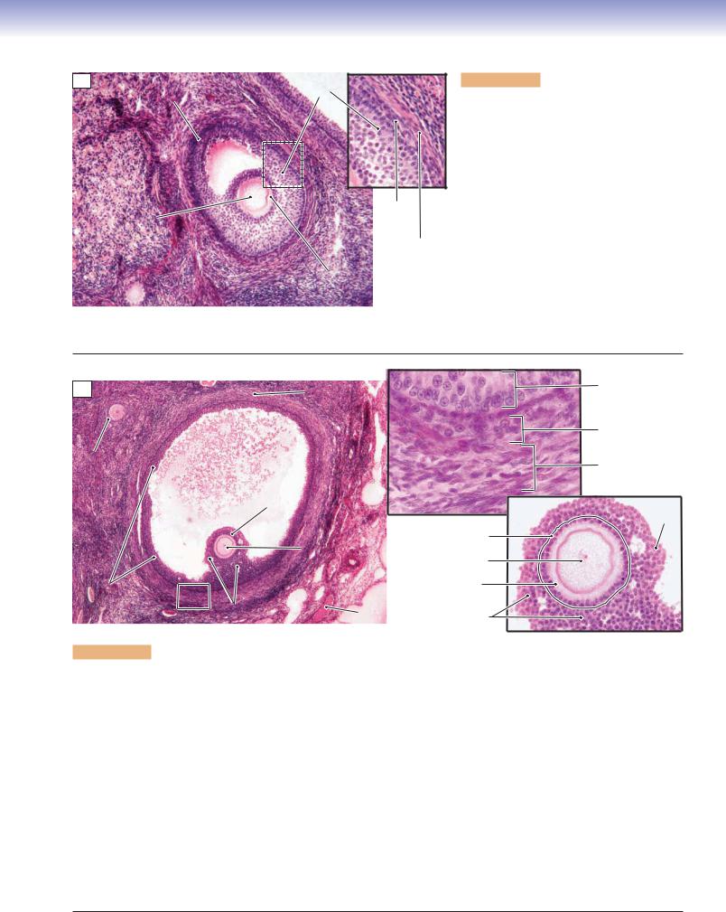
The ovaries are paired organs covered by a simple, usually cuboidal, mesothelium (sometimes called germinal epithelium) and a tunica albuginea (connective tissue). Each ovary is divided into a cortex and a medulla. The cortex contains various stages of follicles including primordial follicles, primary follicles, secondary follicles, and, occasionally, graafian follicles. It may also contain the corpus luteum, a temporary endocrine gland formed by components of an ovulated follicle. A degenerated corpus luteum persists in the ovary as the corpus albicans. Most follicles degenerate (undergo atresia) before ovulation and are then called atretic follicles. The medulla contains connective tissue with blood vessels, nerve fibers, and lymphatic vessels.
Primordial follicle |
Primary follicle (unilaminar) |
Primary follicle (multilaminar) |
|||
|
|
|
|
Zona |
o |
Basement |
Oocyte (1o) |
Basement |
Oocyte (1o) |
pellucida |
Oocyte (1 ) |
membrane |
|
membrane |
|
|
|
D.Cui |
Follicular |
Granulosa |
|
cell (squamous) |
cell (cuboidal) |
Granulosa cell |
|
|
|
|
|
|
Graafian follicle |
Secondary follicle |
|
Theca externa |
|
|
Theca externa |
|
|
|
|
Theca interna |
|
|
|
Granulosa cells |
Antrum |
Antrum |
Theca interna |
|
|
||
Corona radiata |
|
|
Granulosa cells |
(granulosa cells) |
|
|
|
Zona pellucida |
|
|
Zona pellucida |
|
|
|
|
Cumulus |
|
|
Granulosa cells |
|
|
|
|
oophorus |
|
Oocyte (1o) |
D.Cui |
|
|
||
B |
|
Oocyte (1°–2°) |
|
|
|
|
Figure 19-3B. Development of the ovarian follicles.
This illustration shows ovarian follicles from early to late stages: the primordial (resting) follicle, the unilaminar primary follicle, the multilaminar primary (growing) follicle, the secondary (antral or vesicular) follicle, and the graafian (preovulatory) follicle. Each of these follicles contains a primary (10) oocyte, which is an immature ovum. A secondary oocyte is formed shortly before ovulation, when the oocyte completes the first meiotic division. The secondary oocyte does not undergo the second meiotic division unless fertilization occurs. Note that the follicles are not drawn to scale; a graafian (preovulatory) follicle is approximately 1,000 times the diameter of a primordial follicle.
CHAPTER 19 ■ Female Reproductive System |
375 |
A |
|
|
Figure 19-4A. |
Primordial follicles, ovary. H&E, 290; |
|
|
|
|
inset 110 |
|
|
|
|
|
|
||
|
|
|
Primordial follicles are the smallest and most numerous |
||
Squamous |
type of follicles in the cortex of the ovary. Each primordial |
||||
follicle contains a germ cell (primary oocyte) in a resting |
|||||
follicular cells |
|||||
state that may persist for as long as 50 years. The primary |
|||||
|
|
|
|||
|
|
|
oocyte is surrounded by a layer of squamous cells called |
||
|
|
|
follicular cells. These follicular cells are somatic cells that |
||
|
|
|
support the oocyte. The oocyte has a pale appearance |
||
|
|
|
and a large nucleus with a prominent nucleolus. About |
||
|
|
|
|||
|
|
|
1 million follicles are present in the ovaries at the time |
||
|
|
Primordial |
of birth; however, only a few hundred of these follicles |
||
|
|
become mature. The follicles begin to grow at puberty, |
|||
|
|
follicles |
|||
|
|
|
and there is a constant loss of follicles throughout the |
||
|
|
|
reproductive years. At menopause, only a few follicles |
||
|
Primordial |
|
remain. |
||
|
|
|
|
||
|
follicle |
|
|
|
|
|
|
|
|
|
|
B
Mitochondria of
follicle cells
Junction between follicular cells
Follicular
cells
Nucleus of primary oocyte
Basal lamina of follicle
 Nucleus of primary oocyte Mitochondria of primary oocyte
  Granules in oocyte
Nucleus of follicular cell
Nucleus of stromal cell
Figure 19-4B. Primordial follicle. EM, 3,900; inset (color) H&E, 500
The primary oocyte at the center of this primordial follicle may appear to be in interphase of the cell cycle, but it is arrested in dictyotene of prophase I of meiosis. What appear to be patches of heterochromatin in the nucleus are partially decondensed tetrads composed of paired homologous chromosomes. The oocyte has been in prophase of meiosis I before birth of the individual. Follicular cells form a simple squamous epithelium that surrounds the oocyte. Note that these cells adhere tightly to the surface of the oocyte. Indeed, there are junctions between the oocyte and the follicle cells, although they are not readily identifiable here. Neighboring follicular cells are also connected by junctional complexes, and there is a basal lamina between the follicular cells and the surrounding interstitial tissue of the ovarian cortex.

376 UNIT 3 ■ Organ Systems
A |
|
|
Figure 19-5A. |
Primary follicles, ovary. H&E, 202; inset |
|||
|
|
|
438 |
|
|
|
|
|
|
|
|
|
|
||
|
|
|
Primary follicles develop from primordial follicles. Each primary |
||||
|
Germinal |
|
follicle consists of a primary oocyte and cuboidal follicle cells. |
||||
|
|
These follicle cells increase in height (from squamous cells to |
|||||
|
epithelium |
|
|||||
|
|
Primordial |
cuboidal cells), and their cellular layers gradually increase as the |
||||
|
|
follicle |
follicle continues to grow. At this stage, follicle cells are called |
||||
|
|
|
granulosa cells, because their cytoplasm begins to have a granu- |
||||
|
|
|
lar appearance. The primary follicles can be classified into unil- |
||||
|
|
|
aminar primary follicles and multilaminar primary follicles. The |
||||
|
|
Cuboidal follicle cells |
unilaminar primary follicle has a single layer of cuboidal granu- |
||||
|
|
losa cells with a smaller oocyte. The multilaminar primary fol- |
|||||
|
|
of primary |
licle has several layers of cuboidal granulosa cells surrounding a |
||||
|
|
(unilaminar) follicle |
|||||
|
|
relatively large oocyte. As the oocyte increases its size, the zona |
|||||
|
|
|
|||||
|
Zona |
Oocyte |
pellucida emerges as an amorphous layer between the surface |
||||
|
of the oocytes and the surrounding granulosa cells (Fig. 19-5B). |
||||||
pellucida |
|||||||
|
|||||||
|
|
|
Situated outside of the basement membrane of the granulosa |
||||
|
|
|
cells are stromal cells that flatten and develop into a sheath that |
||||
|
|
|
surrounds the follicle; this layer is called the theca folliculi. |
||||
|
|
|
|
|
|
||
Cytoplasm of oocyte |
|
|
|
|
|
||
|
|
|
|
|
|||
|
|
|
|
|
|
|
|
B
Primary |
Theca |
|
oocyte |
folliculi |
Primordial |
Granulosa |
|
follicle |
cells |
|
|
Zona pellucida
Granulosa cells
Microvilli
Theca folliculi
Figure 19-5B. Growing (primary) follicle. EM, 3,200; inset (color) H&E, 152
The oocyte in the center of this growing follicle has been sectioned off center so that the nucleus is not shown. Although the oocyte has begun to grow, it is still arrested in prophase of meiosis I. Note the membrane-bound vesicles in the cytoplasm of the oocyte; these will participate in the cortical granule reaction if the oocyte becomes fertilized. The follicle cells that surrounded the oocyte have proliferated and transformed into granulosa cells. At this stage, the granulosa comprises about two layers of cuboidal cells. The inner granulosa cells no longer have smooth close contact with the surface of the oocyte because a layer of amorphous extracellular material, the zona pellucida, has developed. As the granulosa cells continue to proliferate, several layers of cells will accumulate, and ultimately, a fluid-filled space, the antrum, will develop. Changes are also underway in the stroma adjacent to the growing follicle. The stromal cells (fibroblasts) have become concentrated and flattened against the basal lamina of the granulosa. These theca folliculi cells will develop properties of steroid hormone–synthesizing cells if development of the follicle continues.
CHAPTER 19 ■ Female Reproductive System |
377 |
A
Granulosa Secondary cells follicle
Antrum
Theca Primary interna oocyte
Theca externa
Zona pellucida
Figure 19-6A. Secondary follicles, ovary. H&E,
108; inset 211
The secondary follicle develops from the continued growth of the multilaminar primary follicle. Spaces filled with follicular fluid (liquor folliculi) appear among the granulosa cells within the secondary follicle. These spaces gradually merge to form a single large space called the antrum. The zona pellucida is distinct, and the theca folliculi (surrounding the follicle) develops into the theca interna and theca externa. The theca interna is the inner vascular layer containing cuboidal (steroid-producing) secretory cells. These cells secrete androgens, which diffuse into the granulosa cells where they are converted into estrogens in response to FSH. The theca externa is an outer connective tissue layer containing mainly collagen and some small squamous cells mixed with a few smooth muscle cells.
B
Theca externa |
Granulosa cells |
|
 Theca interna
Theca interna
Primary |
|
|
Theca externa |
follicle |
Antrum |
|
|
|
|
||
|
|
|
|
|
|
Corona |
|
|
|
radiata |
Cumulus |
|
|
|
oophorus |
|
|
Oocyte |
Corona radiata |
|
|
Nucleus of oocyte |
|
|
|
|
|
Membrana granulosa |
|
|
Zona pellucida |
|
|
|
|
(granulosa cells) |
Cumulus |
Blood |
Cumulus oophorus |
|
oophorus |
vessel |
Figure 19-6B. Graafian follicles, ovary. H&E, 54; inset (upper) 429; inset (lower) 178
The Graafian follicle is a mature follicle; it is also called a preovulatory follicle. At this stage, the follicle has grown to a large size (about 25 mm) and bulges from the surface of the ovary. The decreased number of granulosa cells and increased volume of fluid in the antrum result in the oocyte being located at the periphery of the follicle. The membrana granulosa is formed by multiple cellular layers of granulosa cells lining the inner wall of the antrum. Some granulosa cells form a hillock called the cumulus oophorus, which supports and houses the oocyte. The inner granulosa cells of the cumulus oophorus form a single layer called the corona radiata, which immediately surrounds the oocyte. As the follicle grows, most of the granulosa cells gradually loosen from the cumulus oophorus, but the corona radiata remains in contact with the oocyte. Eventually, the oocyte, with the corona radiata, floats freely in the antrum before ovulation. The oocyte remains as a primary oocyte in the graafian follicle until pituitary secretion of LH increases sharply (LH surge); this stimulates the primary oocyte to complete the first meiotic division and become a secondary oocyte. The secondary oocyte with the corona radiata and polar body (from the first oocyte division) are released from the graafian follicle of the ovary. After the secondary oocyte reaches the ampulla of the oviduct, the second meiotic division occurs, if fertilization takes place. A spermatozoan must penetrate the corona radiata and zona pellucida to complete the fertilization process. The upper inset shows the theca folliculi (theca interna and theca externa). The lower inset shows the oocyte surrounded by granulosa cells.
The ovarian cycle is under the control of the hormones FSH and LH produced by the gonadotrophs of the anterior pituitary gland. FSH stimulates estrogen production and follicular growth; LH stimulates meiotic division of the primary oocyte, ovulation, and development of the corpus luteum. The estrogens play an important role in the stimulation of follicle growth by promoting proliferation of the granulosa cells, and they also stimulate the mammary glands to prepare for lactation.

378 UNIT 3 ■ |
Organ Systems |
|
A |
Theca |
Theca |
|
lutein cells |
lutein cells |
Blood |
|
|
vessels |
|
|
Corpus |
|
Ovulated |
albicans |
Corpus luteum |
secondary oocyte |
|
Nuclei of granulosa |
|
Granulosa |
lutein cells |
|
Connective |
||
lutein cells |
||
tissue |
||
|
Cytoplasm of granulosa lutein cells
Figure 19-7A. Corpus luteum, ovary. H&E, 36; insets 363
After ovulation, the remaining portion (wall) of the graafian follicle transforms into the corpus luteum (yellow body). The wall of the corpus luteum is folded and contains granulosa lutein cells (derived from granulosa cells) and theca lutein cells (from the theca interna). The granulosa lutein cells are large and have pale cytoplasm; these cells have features of steroid hormone–producing cells, and they produce primarily progesterone. The theca lutein cells are smaller but also have features of steroid hormone–secreting cells; these cells secrete primarily progesterone and androgens.
B
Blood vessels
Corpus albicans
Corpus albicans
Figure 19-7B. Corpus albicans, ovary. H&E, 34
In the absence of fertilization, the corpus luteum is active only for a short period of time (10–14 days). The corpus luteum degenerates, decreases in size, and forms a structure called the corpus albicans. The corpus albicans consists of dense connective tissue that appears as a white scar; it gradually decreases in size and remains in the ovary for months to years. However, if fertilization and implantation occur, the corpus luteum is rescued from degeneration by human chorionic gonadotropin (hCG) hormone from the placenta. During pregnancy, the corpus luteum will remain active for the first 6 months of gestation, after which it degenerates, and the corpus albicans is formed. Formation of the corpus luteum is stimulated by the LH surge.
CLINICAL CORRELATION
C
Granulosa cell tumor with  neoplastic cells arranged in cords
Figure 19-7C. Granulosa Cell Tumor. H&E, 52
Granulosa cell tumor of the ovary is a neoplasm composed of ovarian granulosa and, occasionally, theca cells. Granulosa cell tumors may arise at any age and are divided into juvenile and adult types. These tumors may produce excess estrogen, the result of which may cause precocious puberty, endometrial hyperplasia, and endometrial cancer. Symptoms may include abdominal pain, hemoperitoneum with hypotension, and mimicking an ectopic pregnancy in younger patients because of rupture of the tumor. Histologically, the tumor cells are small and cuboidal, and may be arranged in a variety of patterns including solid, trabecular, and cordlike. The tumor cells often contain a groove resembling a coffee bean. Small follicle-like structures named Call-Exner bodies may be visible in well-differentiated tumors. The behavior of granulosa cell tumors is variable and may take an aggressive course in some patients. A total abdominal hysterectomy and bilateral salpingo-oophorec- tomy are the treatments of choice in the early stage.

CHAPTER 19 ■ Female Reproductive System |
379 |
Hormone concentration in plasma
|
|
(7) |
|
|
(1) FSH |
LH |
|
|
Pituitary |
|
|
|
|
gonadotroph |
|
|
(9b)Progesterone |
hormones |
|
|
|
|
||
|
(4b) |
|
(11b) |
|
|
|
|
||
|
|
(6) |
Estradioll |
|
|
|
|
|
|
|
|
|
|
Ovarian |
|
|
|
|
steroid |
|
|
|
|
hormones |
|
|
|
(9a) |
|
(3) |
(5) |
(8) |
(10) |
(11a) |
(2) |
|
|
|
Ovary |
|
|
|
|
|
|
(4a) |
|
|
|
|
|
|
|
Endometrium |
0 |
7 |
14 |
21 |
28 |
J. Naftel |
|
|||||
|
Day of nominal female reproductive cycle |
|
|
|
|
Figure 19-8. Events of the female reproductive cycle.
The following sequence of events refer to the numbered events labeled in red in the diagram above. (1) At the beginning of the female reproductive cycle, there are rising levels of gonadotropic hormones from the anterior pituitary, most importantly FSH. (2) This rise promotes ovarian recruitment of a cohort of antral follicles to proceed into advanced development and then selection of typically a single dominant follicle at about day 6. (3) These follicles secrete steroid hormones, most prominently estrogens, that (4a) promote rebuilding of the endometrium (proliferative phase) and (4b) exert a negative feedback on FSH secretion by pituitary gonadotropes.
(5) In the latter part of the follicular phase, the dominant follicle secretes increasing amounts of estrogens (and, to a lesser extent, progesterone). (6) When circulating estrogen reaches a threshold level (about 200 pg/mL) for a duration of about 36 hours, pituitary gonadotropes are stimulated to sharply increase secretion of gonadotropic hormones––most importantly, LH. (7) This LH surge from the pituitary brings about final maturation of the dominant follicle culminating in ovulation (about 40 hours after initiation of the LH surge) and formation of the corpus luteum from the remaining components of the follicle. (8) The corpus luteum secretes progesterone as well as estrogens. (9a) This induces a change in the endometrium from the proliferative phase to the secretory phase. (9b) Meanwhile, gonadotropin secretion is greatly reduced, probably because of negative feedback effects of the high progesterone and estrogen levels coming from the corpus luteum. (10) Without LH support, the corpus luteum fails after about 10 days, and steroid hormone levels fall. (11a) This loss of steroid hormone support results in degenerative changes in the endometrium culminating in menstruation. (11b) The fall in progesterone also releases the pituitary gonadotropes from negative feedback with the result that FSH secretion starts to rise toward the end of the cycle, and this starts another round of follicle recruitment.

380 UNIT 3 ■ Organ Systems
Oviducts (Fallopian Tubes)
A
Ciliated cells
Lumen
Cilia
Mucosa
Muscularis
Lamina propria
Serosa |
Peg cells |
|
Figure 19-9A. Oviduct (fallopian tube). H&E, left 17; right 680
The oviduct (fallopian tube) can be divided into four regions: the infundibulum, ampulla, isthmus, and intramural portion (Fig. 19-1). The infundibulum is a funnel-shaped opening that has a fringe of tentacle-like extensions called fimbriae. The ampulla has a relatively large, labyrinthine lumen where fertilization usually takes place. The isthmus is a narrow portion of the oviduct, close to the uterus. The intramural portion is the terminal segment and is located within the uterine wall. The wall of the oviduct consists of a mucosa (simple columnar epithelium and lamina propria), muscularis (inner circular and outer longitudinal smooth muscle), and serosa. The epithelium of the oviduct contains ciliated cells and peg cells. The cells vary in height according to hormonal stimulation. The oviduct provides an ideal environment for the fertilization of the oocyte and initial development of the embryo as well as transportation of the zygote (fertilized oocyte) to the uterus. On the left is a low-magnification view of the ampulla; on the right is a higher magnification view of the mucosa. Ciliated cells help sweep the oocyte toward the uterus. Each ciliated cell has a pale appearance with many cilia on its apical surface. These cells have a large nucleus and a fair amount of cytoplasm. Peg cells are secretory cells that produce nutrient-rich secretions to nourish and protect the oocyte and promote fertilization. They are small in size and interspersed among the ciliated cells.
B
 Cilia
Cilia
 Microvilli
 Basal bodies
Nucleus of ciliated cell
Figure 19-9B. Epithelial cells lining the oviduct. EM, 8,900
The simple columnar epithelium that lines the oviduct is composed of two cell types (ciliated cells and peg cells); only ciliated cells are shown here. These ciliated cells function, along with smooth muscle of the muscularis, in mixing the contents (gametes) of the lumen and in transporting the oocyte and zygote at a precisely controlled rate along the length of the lumen of the oviduct. The number and activity of cilia change in response to changes in the levels of steroid hormones throughout the reproductive cycle, reaching a peak at the time of ovulation when estrogens dominate. Note that these cells also bear numerous microvilli, suggesting an additional absorptive function.
 Basal lamina

CHAPTER 19 ■ Female Reproductive System |
381 |
Uterus
Uterine
glands
Functionalis 
|
Sloughed |
Endometrium |
gland |
|
|
|
Basalis |
|
Myometrium |
|
|
|
Arteries in the |
A |
Myometrium |
myometrium |
|
|
Figure 19-10A. Menstrual phase of the endometrium, uterus (days 1–4 of the cycle). H&E, 13; insets 93
The wall of the uterus includes the endometrium, the myometrium, and serosa. The endometrium, the mucosa of the uterus, is composed of a surface epithelium and simple tubular uterine glands within a stroma of connective tissue. The endometrium consists of the basalis (basal layer) and the functionalis (functional layer). The functionalis is near the lumen and undergoes changes during the menstrual cycle. During the menstrual phase, the functionalis sloughs off as a result of ischemia and necrosis caused by contraction of the coiled arteries. This occurs when fertilization does not take place and the corpus luteum atrophies, causing the levels of estrogen and progesterone to fall. The menstrual phase is the initial stage of the menstrual cycle; the endometrium will begin to recover at the end of the menstrual phase.
Luminal surface
Endometrium |
|
Straight |
|
|
|
Basalis |
uterine glands |
Straight |
|
|
|
|
||
|
|
uterine glands |
|
|
|
|
|
|
|
|
|
|
Mitotic |
Lumen of |
|
|
|
figures |
gland |
|
Myometrium |
|
|
|
B |
|
|
Glandular epithelium |
|
Figure 19-10B. Proliferative phase of the endometrium, uterus (days 5–14 of the cycle). H&E, 18; inset (upper) 68; inset (lower) 293
The proliferative phase follows the menstrual phase. The epithelium, uterine glands, and connective tissue of the functionalis are rebuilt by proliferation and differentiation of cells that remained in the basalis. At this stage, the uterine glands are straight and have narrow lumens as shown here; the surface of the endometrium is smooth. The epithelial lining of the uterine glands commonly appears as pseudostratified columnar epithelium because of proliferation of the lining cells. Mitotic figures are occasionally seen (inset). The glands open onto the luminal surface of the uterus. During the proliferative phase, the changes in the endometrium are driven by estrogens that are produced by the granulosa cells of the developing follicles.
Luminal |
Lumen of |
|
uterine glands |
||
surface |
||
|
Uterine
glands
Functionalis
Stroma
Endometrium
Coiled arteries
|
Basalis |
|
Myometrium |
Lumen of |
|
uterine gland |
||
|
C
Figure 19-10C. Secretory phase of the endometrium, uterus (days 15–28 of the cycle). H&E, 14; insets 89
The secretory phase begins shortly after ovulation occurs. It is influenced by progesterone produced by the corpus luteum. At this stage, the endometrium becomes thickest (6–7 mm), and the uterine glands are coiled and have large sacculated lumens. The upper inset shows tortuous glands with large, irregular, sawtooth-shaped lumens. The lower inset shows coiled arteries found in the endometrial stroma. These coiled arteries are also called spiral arteries and extend transiently from the basalis into the functionalis of the endometrium. The coiled arteries arise from arcuate arteries of the myometrium. During the secretory phase, these spiral arteries become elongated and highly coiled and extend into the functionalis of the endometrium. The arcuate arteries also give rise to straight arteries that permanently supply the basalis.

382 |
UNIT 3 ■ |
Organ Systems |
|
|
|
|
|
|
|
|
Implantation |
|
|
|
|
|
|
|
Implantation |
|
|
|
|
Blastocyst |
|
|
|
||
|
site |
|
Morula |
|
|
|
|
||
|
|
|
D.Cui |
|
|
|
Amniotic |
|
Blastocyst |
|
|
|
|
|
|
cavity |
|
||
|
|
|
|
|
|
|
|
|
cavity |
|
|
Uterine |
|
Trophoblast |
Inner cell |
|
|
|
|
|
|
glands |
|
|
|
Endometrium |
|||
|
|
Zona |
|
|
mass |
||||
|
|
|
|
|
|||||
|
|
|
pellucida |
|
Blastocyst (embryoblast) |
Syncytiotrophoblast |
|||
|
A |
|
Blastomeres |
|
cavity |
Cytotrophoblast |
Bilaminal |
||
|
|
(subdivided zygote) |
(blastocoel) |
embryonic disc |
|||||
|
|
|
|
|
|||||
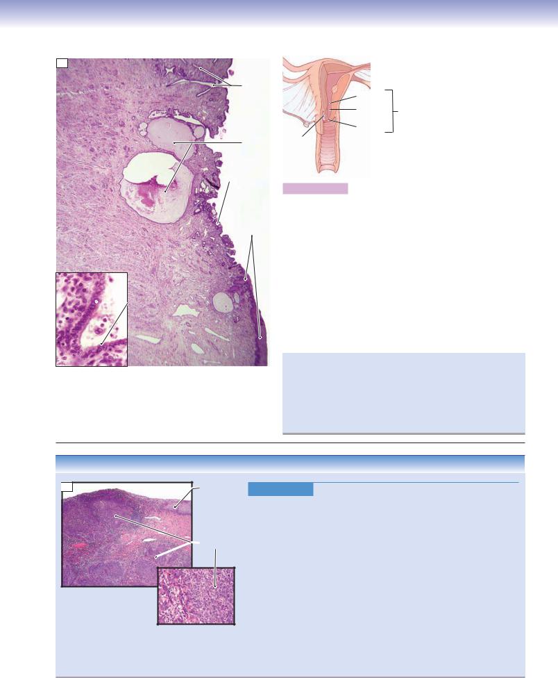
Figure 19-11A. Implantation, endometrium of the uterus. H&E, 8
After an ovum has been successfully fertilized by a spermatozoan in the ampulla of the oviduct, the zygote (fertilized oocyte) undergoes mitotic cell division (cleavage) and becomes a multicellular structure called the morula. The morula develops into the blastocyst, which is transported into the uterus. The process of the blastocyst attaching to the endometrium of the uterus is called implantation. Implantation occurs at the end of the secretory phase; the endometrium during this period of time is also called the premenstrual endometrium (days 25–28). Implantation usually occurs on the posterior wall of the body of the uterus. If implantation succeeds, the trophoblast differentiates into two cell layers: an inner cytotrophoblast layer and an outer syncytiotrophoblast layer. The syncytiotrophoblast attaches to and invades the endometrium of the uterus, and the process of placentation begins. hCG secreted by the placenta stimulates the corpus luteum to remain active and continue to secrete estrogen and progesterone during the pregnancy. The photomicrograph on the left shows an implantation site enclosed within the connective tissue of the endometrium.
CLINICAL CORRELATIONS
B
C
Adenocarcinoma  invading the
myometrium
Figure 19-11B. Endometrial Adenocarcinoma. H&E, 48
Endometrial adenocarcinoma is the most common form of endometrial cancer, accounting for approximately 80% of cases. The majority of cases of endometrial adenocarcinoma arise in the setting of elevated levels of estrogen unopposed by the action of progesterone, causing endometrial hyperplasia. Some cases, however, arise in postmenopausal women with atrophy of the endometrium. Excess or unopposed estrogen may be due to chronic anovulation, obesity, ovarian granulosa cell tumors, or exogenous hormone intake. In the early stage, the cancer is usually asymptomatic. Common symptoms include vaginal bleeding, menorrhagia, metrorrhagia, and lower abdominal pain. Histologically, the cancer is characterized by the presence of cells resembling the glandular cells of the endometrium, and range from well differentiated with gland formation to poorly differentiated with solid sheets of neoplastic cells. Endometrial biopsy is widely used in the diagnosis of the cancer. Treatment options include surgical removal of the uterus, radiation therapy, and chemotherapy.
Fascicles of smooth muscle
Figure 19-11C. Uterine Leiomyoma. H&E, 95
Uterine leiomyoma, or fibroid, is a benign neoplasm, derived from smooth muscle cells of the uterine myometrium. Leiomyomas represent the most common benign neoplasm in women, and occur more frequently in African Americans. Leiomyomas occur in the reproductive years when estrogen levels are high, and tend to regress during menopause. Most patients with fibroids are asymptomatic, but, as the tumor enlarges, symptoms may include abnormal bleeding, menorrhagia, lower abdominal pain, and increased urinary frequency. Grossly, leiomyomas are well circumscribed and may be in subserosal, intramural, or submucosal locations. Leiomyomas can be single but are often multiple and may become quite large. The cut surface is typically white to tan, with a whorled, bulging appearance. Histologically, the tumor cells appear as well-differentiated, spindle-shaped smooth muscle cells, often with increased extracellular matrix, such as collagen, proteoglycan, and fibronectin. Leiomyomas rarely become their malignant counterpart, leiomyosarcomas, which usually develop de novo. Treatment options include hysterectomy, myomectomy (removal of the fibroid), and hormone therapy.
CHAPTER 19 ■ Female Reproductive System |
383 |
A
Branched |
|
cervical |
|
glands |
Internal os |
|
|
|
Endocervical |
|
canal |
|
External os |
Cervical |
Ectocervix |
cysts |
|
Cervix
 EndocervixEndocervix
Stratified squamous
Cervical stroma  epithelium (ectocervix)
Simple columnar  epithelium
(endocervix)
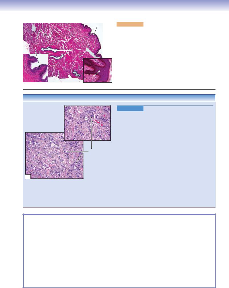
Figure 19-12A. Cervix. H&E, 17; inset 350
The inferior part of the uterus forms the cervical canal, which bulges into the vagina. The internal os is the opening from the endocervical canal to the uterus; the external os is the opening to the vaginal canal. The surface of the endocervix is lined by simple columnar epithelium, which consists of mucus-secreting cells (inset); the ectocervix is lined by stratified squamous epithelium. The cervix contains long branched mucous glands known as cervical glands; when these glands become obstructed they form cervical cysts (nabothian cysts). The secretion of the cervix changes depending on the stage of the menstrual cycle; however, the mucosa of the cervix does not slough off as does the endometrium of the uterus. The cervical stroma is composed of dense connective tissue mixed with a small amount (about 15%) of smooth muscle. Usually, the cervix has a narrow canal; however, during delivery, dilation of the cervix allows the baby to pass through the canal.
The cervical transformation (transition) zone is the area of the cervical mucosa between the original squamocolumnar junction and the restored or new squamocolumnar junction that is formed through the processes of squamous metaplasia and squamous epithelialization. The majority of cervical carcinomas arise in this zone, and it is important that this area be sampled during screening with a Papanicolaou smear.
CLINICAL CORRELATION
 B
B
Squamous
epithelium
Squamous cell  carcinoma
Figure 19-12B. Cervical Cancer. H&E (upper left), 20; (lower right), 115
Cervical cancer is a malignant neoplasm of the uterine cervix, the majority of which are squamous cell carcinomas. Risk factors include the early onset of sexual activity, multiple sexual partners, and exposure to human papillomavirus (HPV). Invasive squamous cell carcinoma is preceded by precursor lesions called cervical intraepithelial neoplasia, in which dysplastic epithelial changes are present. The majority of intraepithelial lesions are related to infection by HPV. The introduction of screening using the Papanicolaou smear, or “Pap” smear, has dramatically reduced the incidence of invasive cervical lesions. Symptoms of cervical cancer include abnormal vaginal bleeding, postcoital bleeding, and vaginal discharge. Histologically, the cancer typically arises in the cervical transformation zone and may show superficial ulceration with endophytic or exophytic growth patterns. The cancer can spread by direct invasion to nearby tissues and organs or metastasize through hematogenous or lymphatic routes. Gardasil, a vaccine against certain HPV types, is used in young women to prevent infection by the virus. Treatment options include surgical removal of the uterus (hysterectomy), radiation therapy, and chemotherapy.
384 UNIT 3 ■ Organ Systems
Intervillousu |
space |
Amniotic |
|
cavity |
|||
Floating |
|
||
|
Chorionic |
||
chorionic |
|
||
villus |
|
plate |
|
|
|
Fetal |
|
|
|
portion |
Fetal blood cells within the chorionic villus
A
Anchoring
villus
Cytotrophoblastic |
|
|
shell |
|
|
|
Maternal portion |
|
Decidua |
(decidua basalis) |
|
|
|
|
basalis |
Decidual cell |
Cells in |
|
||
|
|
|
|
|
cytotrophoblastic shell |
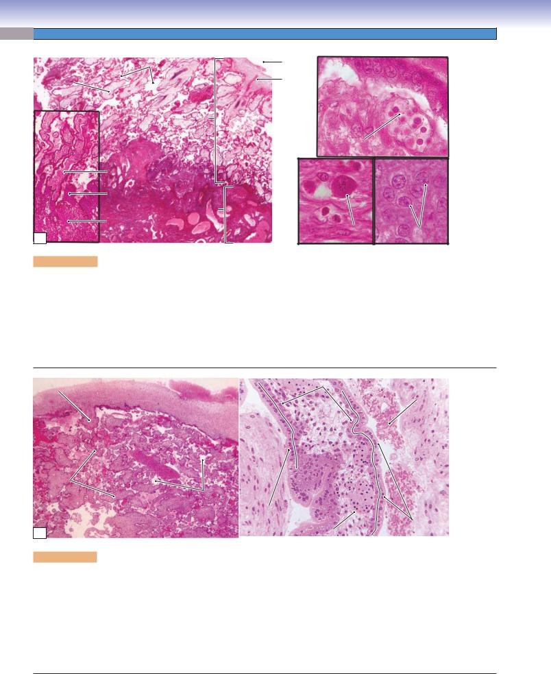
Figure 19-13A. Overview of the placenta. H&E, left 13; left inset 55; right (upper) 704; right (lower) 748
The placenta consists of the maternal portion and the fetal portion. It is a temporary organ that provides a bridge for exchanging gases, nutrients, hormones, and other materials between the maternal and fetal blood circulations. The maternal portion is the decidua basalis. The fetal portion consists of the chorionic plate (Fig. 19-11A), chorionic villi, and cytotrophoblastic shell. Fetal blood flows within the blood vessels of the chorionic villi; maternal blood is contained within the intervillous space. The placental barrier prevents the fetal blood from mixing with the maternal blood. The decidua basalis forms when stromal fibroblasts of the endometrium are transformed into decidual cells at the site of implantation. The syncytiotrophoblast invades the maternal blood vessels replacing smooth muscle in the vessel walls. Syncytiotrophoblasts also line the surface of the intervillous space. The cytotrophoblast forms an interface (cytotrophoblastic shell) between the maternal and fetal tissues.
Stem |
Amniotic cavity |
Cytotrophoblast |
Maternal blood in the |
|
|||
villus |
Chorionic plate |
|
intervillous space |
|
|
|
Chorionic villus
Chorionic
villus
Intervillous
space
Syncytiotrophoblast
Syncytiotrophoblast
B |
Fetal blood vessel |
Figure 19-13B. Fetal portion of the placenta. H&E, left 18; right 136
The chorionic plate consists of connective tissue and forms the wall of the amniotic cavity; it contains chorionic arteries and veins. The chorionic villi can be classified on the basis of their developmental stages: (1) Primary chorionic villi are newly formed villi at an early stage (about the second week of implantation), and consist of only a trophoblast layer. (2) Secondary chorionic villi develop at the end of the second week when mesenchymal tissue grows into the villi and forms a mesenchymal core within the trophoblastic shell. (3) Tertiary chorionic villi develop at the third week, at which time the fetal blood and blood vessels are formed within the chorionic villi. By the end of the third week, the fetal blood begins to flow, and gas and nutrient exchange takes place between the fetal and maternal blood by diffusion through the placental barrier. The placental barrier is composed of the syncytiotrophoblast, cytotrophoblast, connective tissue of the villus, endothelium of the fetal capillary, and the basement membranes of the trophoblast and endothelium. The syncytiotrophoblast produces hCG hormone, which plays an important role in maintaining pregnancy via stimulation of the corpus luteum to secrete progesterone.
CHAPTER 19 ■ Female Reproductive System |
385 |
A
Umbilical
arteries
Mucous connective tissue (Wharton jelly)
Umbilical
vein
Figure 19-14A. Umbilical cord. H&E, 12; inset 79
The umbilical cord is a ropelike structure that connects the developing fetus to the placenta. It contains two umbilical arteries and one umbilical vein. These vessels carry oxygen and nutrients from the mother to the fetus and waste products away from the fetus. The blood vessels are surrounded by a mucous connective tissue (Wharton jelly). The umbilical arteries carry deoxygenated fetal blood to the placenta by way of the chorionic arteries and the chorionic villi. After gas and nutrient exchange with the maternal blood, the oxygenated blood is transported from chorionic veins to the umbilical vein, which returns blood to the fetus.
Funisitis is inflammation of the umbilical cord that often accompanies chorioamnionitis (inflammation of the fetal membranes). Funisitis typically occurs after 20 weeks of gestation, often because of a bacterial infection. Neutrophils migrate through the umbilical vessels and may enter the Wharton jelly. Another possible complication in pregnancy is an umbilical knot. In severe cases, obstruction of blood supply can result in the fetal death.
Vagina
B
|
Lumen |
|
|
Epithelium |
|
|
Epithelium |
|
Mucosa |
Ridges of the |
|
epithelium |
||
Lamina |
||
|
||
propria |
|
|
|
Lamina propria |
|
Muscularis |
Smooth muscle |
|
|
Connective
tissue 
Adventitia
Figure 19-14B. Vagina. H&E, 41; inset (upper) 63; inset (lower) 74
The vagina is a tubular organ that connects the cervix of the uterus to the external genitalia. The wall of the vagina consists of the mucosa, muscularis, and adventitia. The mucosa comprises a nonkeratinized stratified squamous epithelium and an underlying lamina propria
(dense irregular connective tissue with many elastic fibers). The muscularis contains mainly longitudinal smooth muscle and some oblique smooth muscle bundles. The adventitia layer is composed of both dense connective tissue (near the muscularis) and loose connective tissue (outer layer). The vagina is moistened by cervical secretions, and it has many sensory nerve endings in the inferior part near the entrance. The epithelium of the vagina undergoes minimal change during the menstrual cycle. There are numerous elastic fibers in the connective tissue, and ridges (folds) in the mucosa, enabling the vaginal canal to expand during sexual intercourse and during the delivery of a baby.
The Papanicolaou (Pap) smear is a very important diagnostic method used for screening early signs of cervical cancer. Cells from the epithelial surface of the vagina and cervix are collected by using a brush and spatula while the vagina is opened by a speculum. Examination of these sample cells provides valuable information for detecting precancerous changes that may require treatment.

386 UNIT 3 ■ Organ Systems
Mammary Glands
A
Lobe of mammary gland
Lobule of mammary gland
Interlobular
duct
Nipple
Openings 
Lactiferous
duct
Lactiferous
sinus
Skin
Figure 19-15A. Overview of the mammary gland.
|
In humans, there are two multilobed mammary glands, |
|
|
one located within the connective tissue of each breast. |
|
|
These exocrine glands produce milk after a pregnancy. |
|
|
Each gland is composed of 15 to 25 lobes of compound |
|
|
tubuloalveolar glands. Each lobe is separated from others |
|
Pectoralis |
by dense connective tissue and adipose tissue and opens |
|
into a lactiferous duct. The secretory alveoli produce |
||
muscles |
||
|
milk and drain it into the intralobular ducts and then to |
|
|
the interlobular ducts. The interlobular ducts merge into |
|
|
lactiferous sinuses from which the milk empties into the |
|
|
lactiferous ducts (15–25). The female mammary glands |
|
Rib |
begin to enlarge during puberty and undergo changes at |
|
different times based on hormone (estrogen, progester- |
||
|
||
|
one, prolactin, and human placental lactogen) levels. |
B
Adipose tissue
Lobules of
gland 
Myoepithelial cell
Cuboidal epithelial cell
|
Dense irregular |
|
connective |
Adipocytes |
tissue |
Figure 19-15B. Inactive (resting) mammary gland.
H&E, 41; inset 359
An example of a resting mammary gland shows a large amount of dense irregular connective tissue and adipose tissue with small mammary gland lobules. The glandular tissue contains mainly intralobular ducts, which are lined by cuboidal epithelial cells and underlying myoepithelial cells (inset). The resting mammary gland has only a few secretory alveoli, some undeveloped intralobular ducts, interlobular ducts, lactiferous sinuses, and lactiferous ducts.
C
Intralobular
ducts
Interlobular
duct
Alveolus
Lobule of
gland
Alveoli
Figure 19-15C. Active (during pregnancy) mammary gland. H&E, 41
An example of a mammary gland during pregnancy shows large lobules and a relatively small amount of interlobular connective tissue. The glandular tissue contains many proliferated alveoli and intralobular ducts. A large interlobular duct is located within the connective tissue shown here. When the mammary glands begin to secrete milk (lactation), the lumina of the alveoli and the ducts are dilated and filled with milk. The milk contains many lipid droplets and proteins (caseins, lactalbumin, and immunoglobulin A) as well as lactose, ions, vitamins, and water. Secretion of milk is initiated by hormonal changes: decrease of estrogen and progesterone and increase of prolactin after delivery and the loss of the placenta. The milk is released by the milk ejection reflex when stimulated by suckling (Fig. 19-16A).
Connective tissue
CHAPTER 19 ■ Female Reproductive System |
387 |
Smooth |
Skin |
muscle bundles |
|
|
Connective |
|
tissue |
Epithelium of |
|
lactiferous duct |
Lactiferous |
|
ducts |
|
|
stratified squamous |
A |
|
epithelium |
Figure 19-16A. Nipple, mammary gland. H&E, 11; left inset
146; right inset 136
The nipple is a small projection at the center of the breast. It contains 15 to 25 openings of lactiferous ducts within its connective tissue and smooth muscle bundles. It is covered by thin skin and surrounded by the areola (pigmented skin). The nipple has many sensory nerve endings that receive stimulation during suckling. This stimulation results in release of oxytocin from the pars nervosa of the pituitary; the oxytocin stimulates contraction of the myoepithelial cells in the mammary gland. The contraction of the myoepithelial cells pushes milk out of the alveoli and ducts and through the lactiferous ducts to the surface of the nipple. This process is called the milk ejection reflex. The lactiferous ducts shown here are from the proximal portion of the ducts near the lactiferous sinuses.
CLINICAL CORRELATION
Infiltrating duct carcinoma of the breast
B
Figure 19-16B. Adenocarcinoma of the Breast (Breast Cancer).
H&E, left (lower) 44; right (upper) 71
Infiltrating duct carcinoma, or invasive ductal carcinoma, is the most common adenocarcinoma of the breast (breast cancer); it contains no features to further classify it into special types of breast carcinoma, such as lobular, tubular, and mucinous carcinomas. Risk factors for the development of breast cancer include female gender, increasing age, family history, long reproductive life, nulliparity, and the presence of proliferative breast lesions or ductal hyperplasia. Approximately 5% of breast cancers are related to specific gene mutations, including BRCA1 and BRCA2. Common signs and symptoms include a palpable breast mass, bloody discharge from the nipple, change in size or shape of a breast, skin dimpling, inverted nipple, peeling of the nipple skin, and redness or pitting of the skin over the breast. Mammograms and breast exams are used to screen for breast cancer. Biopsy is performed on suspicious lesions to determine a tissue diagnosis. Histologically, breast cancer varies from well-formed glandular structures to sheets of poorly differentiated cells. Histologic grading of breast cancer is based on tubule formation, nuclear pleomorphism, and the mitotic rate. Treatment includes surgical removal of a tumor (lumpectomy), removal of the entire breast (mastectomy) and lymph nodes, radiation therapy, chemotherapy, and hormone therapy.
SYNOPSIS 19 - 1 Clinical and Pathological Terms for the Female Reproductive System
■Endophytic: Term to describe an inward-growing process such as a neoplasm that grows on the interior of an organ (Fig. 19-12B).
■Exophytic: Term to describe an outward-growing process such as a neoplasm that grows externally on an organ or within the lumen of an organ (Fig. 19-12B).
■Hemoperitoneum: Blood within the peritoneal cavity because of a variety of causes including trauma, rupture of a tumor, or rupture of an ectopic pregnancy (Fig. 19-7C).
■Menorragia: Refers to excessive or prolonged uterine bleeding at regular intervals during menstruation; some causes include uterine leiomyomas, anovulation and ovarian dysfunction, hormonal imbalance, bleeding tendency, and malignancy (Fig. 19-11B).
■Metrorragia: Refers to uterine bleeding at irregular intervals often at times between the expected menstrual periods; causes may be similar to menorrhagia and include hormonal imbalance, malignancy, uterine polyps, and bleeding tendency (Fig. 19-11B).
■Nulliparity: Term used to describe never having given birth to a child; by contrast, a woman who has given birth to two or more children is termed multiparous (Fig. 19-16B).

20 Eye
Introduction and Key Concepts for the Eye
Figure 20-1 |
Overview of the Eye |
Figure 20-2 |
Orientation of Detailed Eye Illustrations |
The Eyelids |
|
Figure 20-3A |
Overview of the Upper Eyelid |
Figure 20-3B |
A Representation of the Upper Eyelid, Glands, and Muscles that Control Eyelid |
|
Movements |
Figure 20-3C |
Clinical Correlation: Chalazion |
Figure 20-4A |
Upper Eyelid (Lower Part), Glands of the Eyelid |
Figure 20-4B |
Upper Eyelid (Middle Part) |
Figure 20-4C |
Upper Eyelid (Upper Part), Muscular Control of Upper Eyelid Movement |
Tunica Fibrosa (Tunica Externa) |
|
Figure 20-5A,B |
Cornea |
Figure 20-5C |
Clinical Correlation: LASEK |
Figure 20-6A |
Pavement Epithelium (Anterior Epithelium) |
Figure 20-6B |
Corneal Endothelium (Posterior Corneal Epithelium) |
Refractive Media of the Eye |
|
Figure 20-7A |
Overview of the Lens |
Figure 20-7B |
The Anterior Region of the Lens |
Figure 20-7C |
The Posterior Region of the Lens |
Figure 20-7D |
The Equatorial Region of the Lens |
Figure 20-8A |
Lens and Zonular Fibers |
Figure 20-8B |
A Representation of the Lens and its Function |
Figure 20-8C |
Clinical Correlation: Cataract |
388